. 2014 Jan;39(1):98-106.
doi: 10.1111/ejn.12421. Epub 2013 Nov 18.

Vesicular glutamate transporter 2 is required for the respiratory and parasympathetic activation produced by optogenetic stimulation of catecholaminergic neurons in the rostral ventrolateral medulla of mice in vivo

Affiliations

Vesicular glutamate transporter 2 is required for the respiratory and parasympathetic activation produced by optogenetic stimulation of catecholaminergic neurons in the rostral ventrolateral medulla of mice in vivo

Stephen B G Abbott et al. Eur J Neurosci. 2014 Jan.

Abstract

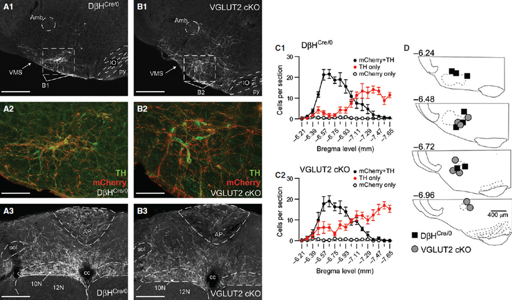
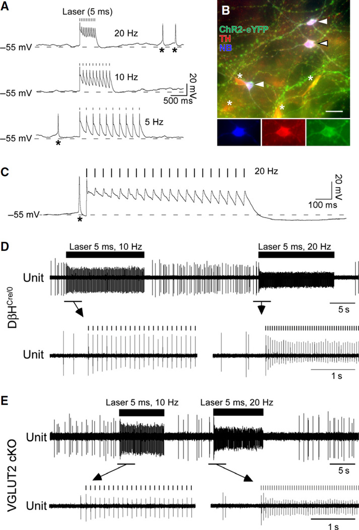
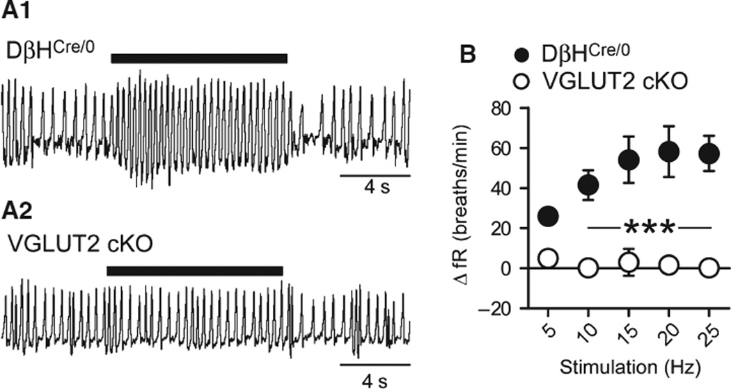
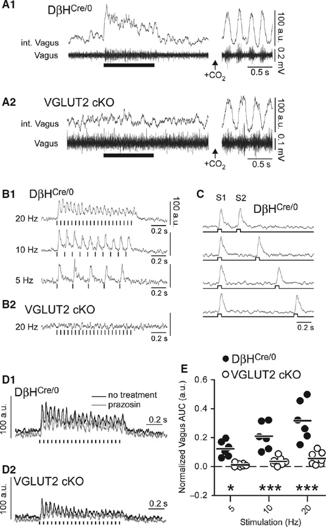
Catecholaminergic neurons of the rostral ventrolateral medulla (RVLM-CA neurons; C1 neurons) contribute to the sympathetic, parasympathetic and neuroendocrine responses elicited by physical stressors such as hypotension, hypoxia, hypoglycemia, and infection. Most RVLM-CA neurons express vesicular glutamate transporter (VGLUT)2, and may use glutamate as a ionotropic transmitter, but the importance of this mode of transmission in vivo is uncertain. To address this question, we genetically deleted VGLUT2 from dopamine-β-hydroxylase-expressing neurons in mice [DβH(Cre/0) ;VGLUT2(flox/flox) mice (cKO mice)]. We compared the in vivo effects of selectively stimulating RVLM-CA neurons in cKO vs. control mice (DβH(Cre/0) ), using channelrhodopsin-2 (ChR2-mCherry) optogenetics. ChR2-mCherry was expressed by similar numbers of rostral ventrolateral medulla (RVLM) neurons in each strain (~400 neurons), with identical selectivity for catecholaminergic neurons (90-99% colocalisation with tyrosine hydroxylase). RVLM-CA neurons had similar morphology and axonal projections in DβH(Cre/0) and cKO mice. Under urethane anesthesia, photostimulation produced a similar pattern of activation of presumptive ChR2-positive RVLM-CA neurons in DβH(Cre/0) and cKO mice. Photostimulation in conscious mice produced frequency-dependent respiratory activation in DβH(Cre/0) mice but no effect in cKO mice. Similarly, photostimulation under urethane anesthesia strongly activated efferent vagal nerve activity in DβH(Cre/0) mice only. Vagal responses were unaffected by α1 -adrenoreceptor blockade. In conclusion, two responses evoked by RVLM-CA neuron stimulation in vivo require the expression of VGLUT2 by these neurons, suggesting that the acute autonomic responses driven by RVLM-CA neurons are mediated by glutamate.

Keywords: C1 neuron; channelrhodopsin 2; conditional knockout; gene disruption in mice; glutamate.


Figures

Fig. 1
Fig. 1
Distribution of ChR2–mCherry-positive neurons in the RVLM, and placement of fiber optics in DβHCre/0 and cKO mice. (A1) Low-magnification image of ChR2–mCherry expression in a coronal section of the RVLM from a DβHCre/0 mouse (approximately −6.64 mm from bregma). Scale bar: 400 µm. (A2) Higher magnification of the region outlined in A1, showing immunofluorescence for ChR2–mCherry (in red) and TH (in green). Colocalisation of ChR2– mCherry and TH is indicated by orange. Scale bar: 100 µm. (A3) ChR2–mCherry-immunoreactive axons within the dorsal medulla of a DβHCre/0 mouse (approximately −7.48 mm from bregma). Scale bar: 200 µm. (B1–B3) Descriptions and scale bars are the same as in A1–A3, except that sections are from a cKO mouse. (C1 and C2) Rostrocaudal distribution of neurons expressing ChR2–mCherry, TH or both in the ipsilateral RVLM of DβHCre/0 (C1, N = 8) and cKO (C2, N = 8) mice. (D) Locations of the tips of implanted fiber optics in DβHCre/0 and cKO mice. 10N, dorsal motor nucleus of the vagus; 12N, hypoglossal motor nucleus; Amb, compact formation of the nucleus ambiguous; AP, area postrema; cc, central canal; IO, inferior olive; py, pyramidal tract; sol, solitary tract; VMS, ventral medullary surface.
Fig. 2
Fig. 2
Characteristics of the response of ChR2–expressing neurons to light stimulation in DβHCre/0 and cKO mice. (A) Current-clamp recording of a ChR2–eYFP-positive RVLM neuron in a slice (DβHCre/0 mouse) during 20-Hz, 10-Hz and 5-Hz stimulation (10 × 5-ms pulses). Each light pulse is followed by a short-latency depolarisation and an action potential. Asterisks indicate spontaneous action potentials. (B) Example of biocytin-filled RVLM-CA neurons that were photo-responsive (arrowheads). All three neurons expressed ChR2–eYFP and TH. Asterisks indicate CHR2–eYFP-positive, TH-positive neurons not labeled with biotin. Inserts correspond to the middle cell in the main panel (black-rimmed arrowhead), and show immunofluorescence for each marker separately. (C) Response of a ChR2–eFYP-positive RVLM-CA neuron to a 1-s stimulus train (20 Hz with 5-ms pulses), illustrating the reduction in action potential amplitude after the first light-induced spike and the hyperpolarisation that follows the stimulus train. (D) Extracellular recording of a single putative ChR2–mCherry-positive RVLM-CA neuron during photostimulation (10 Hz and 20 Hz with 5-ms pulses) in an anesthetised DβHCre/0 mouse. Expanded traces (below) show the pulse-by-pulse activation typical of ChR2-expressing neurons, and the gradual reduction in spike amplitude during stimulation and the silent period following the stimulus train. (E) Identical experiment in a cKO mouse.
Fig. 3
Fig. 3
Effect of stimulating RVLM-CA neurons on fR in DβHCre/0 and cKO mice. (A) Original plethysmography traces (inspiration upward) of a DβHCre/0 mouse (upper trace) and a cKO mouse (lower trace) during photostimulation (5-ms pulses at 20 Hz, 10-s train). (B) Relationship between photostimulus frequency and increase in fR in DβHCre/0 (N = 7) and cKO (N = 7) mice. ***P < 0.001 for DβHCre/0 vs. cKO (Bonferroni post hoc test following repeated measures anova).
Fig. 4
Fig. 4
Effect of stimulating RVLM-CA neurons on efferent vagus nerve activity in DβHCre/0 and cKO mice. (A1 and A2) Left panel: integrated (upper trace) and raw (lower trace) recording of multi-unit vagus nerve activity in urethane-anesthetised DβHCre/0 (A1) and cKO (B2) mice, showing activation by photostimulation (5-ms pulses at 20 Hz, 1-s train). Right panel: respiratory-related oscillations in vagus nerve activity caused by hypercapnia in the same mouse (7% CO2). The amplitude of oscillations in vagus activity under conditions of elevated inspired CO2 (i.e. peak to trough) was assigned a value of 100 a.u. to normalise recordings between experiments. Integrated vagus nerve activity is rectified and smoothed with a 0.03-s time constant. (B1 and B2) Normalised waveform averages of the vagal response to phot-ostimulation (5-ms pulses, 15–20 trials per trace, averaging triggered from the onset of the 1-s stimulus train) in a DβHCre/0 mouse (B1) and a cKO mouse (B2). (C) Waveform average of the vagal response to paired-pulse stimulation (50-ms pulses, identified by S1 and S2) in a DβHCre/0 mouse; note that the magnitude of the second burst was similar regardless of the delay of the second pulse of light down to 250 ms. (D) Example of the of the vagal response to photostimulation (5-ms pulses at 20 Hz) in a DβHCre/0 mouse (D1) and a cKO (D2) mouse before and after α1-adrenoreceptor blockade (prazosin, 1 mg/kg). Waveforms were generated by averaging 10– 15 trials per trace. Note that the cKO mouse used in the example had the largest response of all cKO mice tested. (E) Group data of the normalised vagus nerve activity during photostimulation (see Materials and methods) in DβHCre/0 (N = 6) and cKO (N = 7) mice. *P < 0.05 and ***P < 0.001 for DβHCre/0 vs. cKO by the Bonferroni post hoc test.

Comment in

References

    1. Abbott SB, Stornetta RL, Socolovsky CS, West GH, Guyenet PG. Photostimulation of channelrhodopsin-2 expressing ventrolateral medullary neurons increases sympathetic nerve activity and blood pressure in rats. J. Physiol. 2009;587:5613–5631. - PMC - PubMed
    1. Abbott SB, Kanbar R, Bochorishvili G, Coates MB, Stornetta RL, Guyenet PG. C1 neurons excite locus coeruleus and A5 noradren-ergic neurons along with sympathetic outflow in rats. J. Physiol. 2012;590:2897–2915. - PMC - PubMed
    1. Abbott SB, Depuy SD, Nguyen T, Coates MB, Stornetta RL, Guyenet PG. Selective optogenetic activation of rostral ventrolat-eral medullary catecholaminergic neurons produces cardiorespiratory stimulation in conscious mice. J. Neurosci. 2013;33:3164–3177. - PMC - PubMed
    1. Agassandian K, Shan Z, Raizada M, Sved AF, Card JP. C1 catecholamine neurons form local circuit synaptic connections within the rostroventrolateral medulla of rat. Neuroscience. 2012;227:247–259. - PMC - PubMed
    1. Bellocchio EE, Reimer RJ, Fremeau RT, Jr, Edwards RH. Uptake of glutamate into synaptic vesicles by an inorganic phosphate transporter. Science. 2000;289:957–960. - PubMed

Publication types

MeSH terms

LinkOut - more resources