. 2014 Jan;18(1):115-24.
doi: 10.1111/jcmm.12170. Epub 2013 Nov 17.

Enhanced expression of the central survival of motor neuron (SMN) protein during the pathogenesis of osteoarthritis

Affiliations

Enhanced expression of the central survival of motor neuron (SMN) protein during the pathogenesis of osteoarthritis

Magali Cucchiarini et al. J Cell Mol Med. 2014 Jan.

Abstract

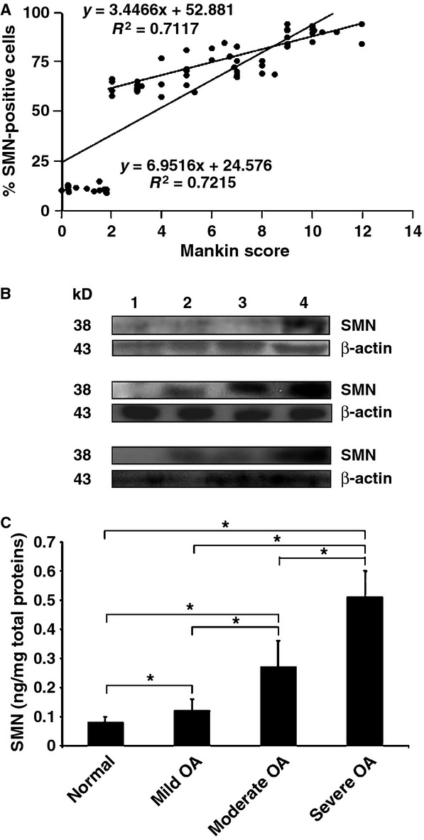
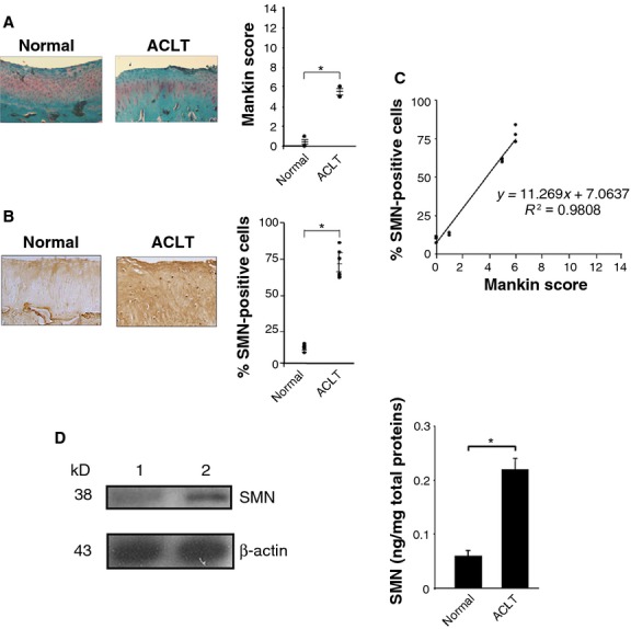
The identification of new components implicated in the pathogenesis of osteoarthritis (OA) might improve our understanding of the disease process. Here, we investigated the levels of the survival of motor neuron (SMN) expression in OA cartilage considering the fundamental role of the SMN protein in cell survival and its involvement in other stress-associated pathologies. We report that SMN expression is up-regulated in human OA compared with normal cartilage, showing a strong correlation with the disease severity, a result confirmed in vivo in an experimental model of the disease. We further show that the prominent inflammatory cytokines (IL-1β, TNF-α) are critical inducers of SMN expression. This is in marked contrast with the reported impaired levels of SMN in spinal muscular atrophy, a single inherited neuromuscular disorder characterized by mutations in the smn gene whereas OA is a complex disease with multiple aetiologies. While the precise functions of SMN during OA remain to be elucidated, the conclusions of this study shed light on a novel pathophysiological pathway involved in the progression of OA, potentially offering new targets for therapy.

Keywords: SMN expression; osteoarthritis progression; pro-inflammatory cytokines.


Figures

Figure 1
Figure 1
Histological grading and survival of motor neuron (SMN) expression in human normal and osteoarthritis (OA) cartilage. All samples were processed for the analysis. (A) Safranin O staining of sections from cartilage explants (representative samples; magnification ×4). (B) Grading of OA severity on safranin O-stained histological sections from all individual explants (normal donors, n = 13, Mankin score 0–2; mild OA patients, n = 14, Mankin score 2–4; moderate OA patients, n = 22, Mankin score 5–8; severe OA patients, n = 16, Mankin score 9–14). (C) SMN-specific immunoreactivity on histological sections from cartilage explants (representative samples; magnification ×10). (D) Percentages of SMN-positive cells on immunohistochemical sections from all individual explants (8.8–14.5%, 57.6–77.0%, 59.6–85.1% and 83.3–94.5% in normal, mild, moderate and severe OA samples, respectively). *Statistically significant difference between groups.
Figure 2
Figure 2
Correlation and biochemical analyses. (A) All individual values of (Fig. 1B and D) were combined to evaluate the correlation between the two parameters. (B) Western blotting analysis in human normal and osteoarthritis (OA) cartilage (1: normal cartilage, 2: mild OA, 3: moderate OA, 4: severe OA; representative samples). (C) Survival of motor neuron concentrations in freshly isolated human normal and OA chondrocytes from all the samples (0.08 ± 0.02, 0.11 ± 0.02, 0.26 ± 0.07 and 0.52 ± 0.06 ng/mg total proteins in normal, mild, moderate and severe OA samples, respectively). *Statistically significant difference between groups.
Figure 3
Figure 3
Molecular analyses. All the samples were processed for the analyses. (A) Survival of motor neuron (SMN)-specific immunoreactivity (representative samples; normal chondrocytes are also shown in regular light; magnification ×200). (B) Abundance of SMN-specific bodies. (C) Correlation between the abundance of SMN-specific bodies and osteoarthritis (OA) severity. The values of Figures 1B and 3B were combined to evaluate the correlation between the two parameters. *Statistically significant difference between groups. (D) Normalized mRNA expression of SMN (1.17–1.22, 1.24–1.27, 1.27–1.33 and 1.32–1.38 in normal, mild, moderate and severe OA samples, respectively) and type-II collagen (1.57–1.59, 1.72–1.76, 1.84–1.88 and 1.91–1.94 in normal, mild, moderate and severe OA samples, respectively). Statistically significant difference between groups for *SMN and +type-II collagen.
Figure 4
Figure 4
Effects of IL-1β and TNF-α upon the survival of motor neuron (SMN) expression levels in human cartilage and chondrocytes. All human normal cartilage explants and cells freshly isolated from these explants were treated for 10 days with either IL-1β or TNF-α (each at either 10 or 100 ng/ml) and processed as described in the Materials and Methods. Explants and cells defined with severe osteoarthritis (OA) were used as positive controls. (A) SMN-specific immunoreactivity on histological explant sections (representative samples; magnification ×20). (B). Safranin O staining of histological explant sections (representative samples; magnification ×2). (C) Percentages of SMN-positive cells on immunohistochemical explant sections (all samples were processed for the analysis) (normal samples: 9.1–12.7% raising at 60.2–63.1%, 73.4–76.8%, 50.8–53.4% and 77.4–78.6% with IL-1β or TNF-α at 10 or 100 ng/ml, respectively; severe OA samples: 84.1–86.4%). (D) SMN concentrations in freshly isolated chondrocytes (all samples were processed for the analysis) (normal samples: 0.10 ± 0.01 ng/mg total proteins raising at 0.18 ± 0.01, 0.22 ± 0.02, 0.14 ± 0.01 and 0.23 ± 0.01 ng/mg total proteins with IL-1β or TNF-α at 10 or 100 ng/ml; severe OA samples: 0.47 ± 0.01 ng/mg total proteins). *Statistically significant difference between groups.
Figure 5
Figure 5
Survival of motor neuron (SMN) expression levels in an experimental osteoarthritis (OA) model in vivo. (A) Safranin O staining of histological sections from rabbit knee joint cartilage (representative samples; view of the femoral condyle; magnification ×4) and grading of OA severity (all samples were processed for the analyses). (B) SMN-specific immunoreactivity on sections from rabbit knee joint cartilage (representative samples; view of the femoral condyle; magnification ×20) and percentages of SMN-positive cells (all samples were processed for the analyses) (normal cartilage: 7.1–13.7%; OA cartilage: 59.7–83.8% as for moderate human OA). (C) Correlation between the SMN expression levels and OA severity (combination of all values from A and B). (D) Analysis of SMN by Western blotting (lane 1, normal cartilage; lane 2, ACLT cartilage; all representative samples) and ELISA (SMN concentrations in all freshly isolated normal and ACLT chondrocytes; *statistically significant difference).

References

    1. Loeser RF, Goldring SR, Scanzello CR, et al. Osteoarthritis: a disease of the joint as an organ. Arthritis Rheum. 2012;64:1697–707. - PMC - PubMed
    1. Poole AR. An introduction to the pathophysiology of osteoarthritis. Front Biosci. 1999;4:D662–70. - PubMed
    1. Goldring MB, Goldring SR. Osteoarthritis. J Cell Physiol. 2007;213:626–34. - PubMed
    1. Lotz M, Loeser RF. Effects of aging on articular cartilage homeostasis. Bone. 2012;51:241–8. - PMC - PubMed
    1. Loughlin J. Genetics of osteoarthritis. Curr Opin Rheumatol. 2011;23:479–83. - PubMed

Publication types

Substances