. 2013 Nov 15:13:544.
doi: 10.1186/1471-2334-13-544.

Humoral and cellular responses to a non-adjuvanted monovalent H1N1 pandemic influenza vaccine in hospital employees

Affiliations

Humoral and cellular responses to a non-adjuvanted monovalent H1N1 pandemic influenza vaccine in hospital employees

Ma Teresa Herrera et al. BMC Infect Dis. .

Abstract

Background: The efficacy of the H1N1 influenza vaccine relies on the induction of both humoral and cellular responses. This study evaluated the humoral and cellular responses to a monovalent non-adjuvanted pandemic influenza A/H1N1 vaccine in occupationally exposed subjects who were previously vaccinated with a seasonal vaccine.

Methods: Sixty healthy workers from a respiratory disease hospital were recruited. Sera and peripheral blood mononuclear cells (PBMCs) were obtained prior to and 1 month after vaccination with a non-adjuvanted monovalent 2009 H1N1 vaccine (Influenza A (H1N1) 2009 Monovalent Vaccine Panenza, Sanofi Pasteur). Antibody titers against the pandemic A/H1N1 influenza virus were measured via hemagglutination inhibition (HI) and microneutralization assays. Antibodies against the seasonal HA1 were assessed by ELISA. The frequency of IFN-γ-producing cells as well as CD4+ and CD8+ T cell proliferation specific to the pandemic virus A/H1N peptides, seasonal H1N1 peptides and seasonal H3N2 peptides were assessed using ELISPOT and flow cytometry.

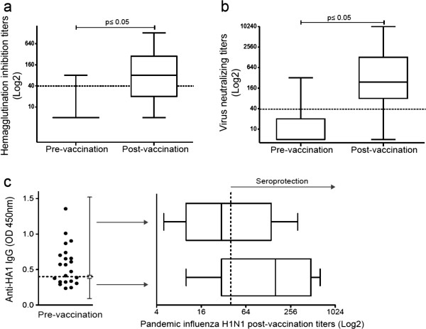
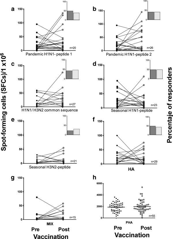
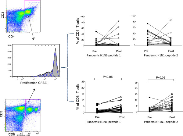
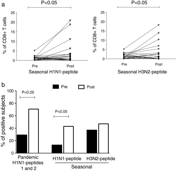
Results: At baseline, 6.7% of the subjects had seroprotective antibody titers. The seroconversion rate was 48.3%, and the seroprotection rate was 66.7%. The geometric mean titers (GMTs) were significantly increased (from 6.8 to 64.9, p < 0.05). Forty-nine percent of the subjects had basal levels of specific IFN-γ-producing T cells to the pandemic A/H1N1 peptides that were unchanged post-vaccination. CD4+ T cell proliferation in response to specific pandemic A/H1N1 virus peptides was also unchanged; in contrast, the antigen-specific proliferation of CD8+ T cells significantly increased post-vaccination.

Conclusion: Our results indicate that a cellular immune response that is cross-reactive to pandemic influenza antigens may be present in populations exposed to the circulating seasonal influenza virus prior to pandemic or seasonal vaccination. Additionally, we found that the pandemic vaccine induced a significant increase in CD8+ T cell proliferation.


Figures

Figure 1
Figure 1
Increased humoral responses after pandemic influenza A/H1N1 vaccination. Specific antibodies against the pandemic influenza virus A/H1N1 were measured via (a) hemagglutination inhibition and (b) microneutralization. The box plots show quartiles and medians. The dotted lines indicate a cut-off value of 1:40, with p ≤ 0.05 (pre-vaccination vs. post-vaccination), as determined using the Wilcoxon singed rank test (n = 60). (c) The presence of IgG antibodies against hemagglutinin A/H1N1 (HA1) was assessed in sera obtained prior to H1N1 vaccination using ELISA, with the dotted line indicating the cut-off value (n = 22). The antibody titers against the pandemic influenza virus A/H1N1 were assessed post-vaccination in sera from individuals with (n = 13) or without (n = 9) anti-HA1 antibodies prior to vaccination using the hemagglutination inhibition assay. The depicted box plots show the quartiles and medians. The dotted line indicates the protective cut-off value.
Figure 2
Figure 2
Frequency of IFNγ-producing cells in response to pandemic influenza peptides. Pre- and post-vaccination PBMCs were stimulated with 10 μg/ml of pandemic H1N1 peptides 1 and 2, a common H1N1/H3N2 sequence, the seasonal H1N1 and H3N2 peptides (a-e), the whole HA protein (f), a mixture of all the peptides (g) or PHA (h) for 48 h. Cells producing IFN-γ were enumerated using an ex-vivo ELISPOT assay. The results are expressed as the number of SFCs per 106 PBMCs. Insets depict the percentage of subjects with a positive IFN-γ response pre- (dark) and post-(light) vaccination.
Figure 3
Figure 3
Proliferative T cell responses to pandemic H1N1 and seasonal peptides. CFSE-labeled PBMCs (1 × 10⁶) were stimulated with 10 μg/ml of pandemic A/H1N1 peptides 1 and 2 for 7 days. The cells were stained with anti-human CD3PE-Texas Red, CD8PE-Cy7 and CD4APC-Cy7 and then analyzed using flow cytometry. The plots show the percentages of proliferating CD4+ or CD8+ T cells prior to and after A/H1N1 vaccination (n = 32).
Figure 4
Figure 4
Pandemic H1N1 vaccination induced CD8+ T cell proliferation to pandemic H1N1, seasonal H1N1 and H3N2 peptides. CFSE-labeled PBMCs (1 × 10⁶) were stimulated with 10 μg/ml of the pandemic H1N1 and seasonal H1N1 and H3N2 peptides for 7 days. The cells were harvested and stained with anti-human CD3PE-Texas Red and CD8PE-Cy7 and analyzed using flow cytometry. (a) Percentages of the CD8+ T cell proliferation pre-vaccination and post-vaccination in response to seasonal H1N1 and H3N2 peptides. The means and standard errors are shown, with p ≤ 0.05 pre-vaccination vs. post-vaccination, according to the Wilcoxon matched-pairs signed rank test. (b) Percentages of positive subjects responding to the pandemic H1N1 peptides 1 and 2 (p < 0.05) and the seasonal influenza A H1N1 (p < 0.05) and H3N2 serotypes post-vaccination (p > 0.05).

References

    1. Chowell G, Bertozzi SM, Colchero MA, Lopez-Gatell H, Alpuche-Aranda C, Hernandez M, Miller MA. Severe respiratory disease concurrent with the circulation of H1N1 influenza. N Engl J Med. 2009;361(7):674–679. doi: 10.1056/NEJMoa0904023. - DOI - PubMed
    1. Greenberg ME, Lai MH, Hartel GF, Wichems CH, Gittleson C, Bennet J, Dawson G, Hu W, Leggio C, Washington D. et al.Response to a monovalent 2009 influenza A (H1N1) vaccine. N Engl J Med. 2009;361(25):2405–2413. doi: 10.1056/NEJMoa0907413. - DOI - PubMed
    1. Nolan T, McVernon J, Skeljo M, Richmond P, Wadia U, Lambert S, Nissen M, Marshall H, Booy R, Heron L. et al.Immunogenicity of a monovalent 2009 influenza A(H1N1) vaccine in infants and children: a randomized trial. JAMA. 2010;303(1):37–46. doi: 10.1001/jama.2009.1911. - DOI - PubMed
    1. Wu J, Li W, Wang HQ, Chen JT, Lv M, Zhou JC, Liang XF, Fang HH, Liu Y, Liu LY. et al.A rapid immune response to 2009 influenza A(H1N1) vaccines in adults: a randomized, double-blind, controlled trial. J Infect Dis. 2010;202(5):675–680. doi: 10.1086/655226. - DOI - PubMed
    1. Virus strains recommended by the World Health Organization for the development of the influenza A (H1N1) vaccines. http://www.who.int/csr/resources/publications/swineflu/2009_05_27_X179Aa....

Publication types