Review
. 2013 Nov 14;39(5):806-18.
doi: 10.1016/j.immuni.2013.10.010.

The spleen in local and systemic regulation of immunity

Affiliations
Review

The spleen in local and systemic regulation of immunity

Vincenzo Bronte et al. Immunity. .

Erratum in

Abstract

The spleen is the main filter for blood-borne pathogens and antigens, as well as a key organ for iron metabolism and erythrocyte homeostasis. Also, immune and hematopoietic functions have been recently unveiled for the mouse spleen, suggesting additional roles for this secondary lymphoid organ. Here we discuss the integration of the spleen in the regulation of immune responses locally and in the whole body and present the relevance of findings for our understanding of inflammatory and degenerative diseases and their treatments. We consider whether equivalent activities in humans are known, as well as initial therapeutic attempts to target the spleen for modulating innate and adaptive immunity.


Figures

Figure 1
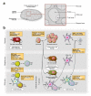
Figure 1. Origins, behavioral activities and functions of splenic immune cell subsets
A. Schematic view of spleen’s anatomy. B. The cartoon depicts the location of several innate and adaptive immune cell components that are found in the resting spleen and can be involved in disease. The orange boxes identify the cells’ origins or the mechanisms that control their positioning or motility within the spleen. The white boxes identify generic functions attributed to the splenic immune cell subsets. Ag, antigen; CDP, Common DC progenitor; CR2, cannabinoid receptor 2; DC, dendritic cell; FDC, follicular DC; FO B cell, follicular B cell; GRK2, guanine nucleotide–binding protein–coupled receptor kinase-2; LTβ, lymphotoxin beta; LXR, liver X receptor; MZ, marginal zone; RA, retinoic acid, S1P1, sphingosine-1 phosphate-1. See also the Immunology image resource (http://www.cell.com/immunity/image_resource-spleen), which provides a collection of images of the spleen and its cellular constituents.
Figure 2
Figure 2. Splenic myeloid cell production and mobilization under inflammatory conditions
In the steady-state, circulating HSPCs do not seed the spleen and can re-engraft the bone marrow. Several inflammatory conditions including some cancers, myocardial infarction and atherosclerosis, however, induce HSPC survival and engraftment in the spleen, followed by local monocytic and granulocytic cell production. Some of the newly made cells may remain in their native tissue where they participate in the regulation of immune tolerance (see Fig 3) or relocate to distant tissues. The orange boxes identify molecular mechanisms that are known to orchestrate these sequential processes. AngII, angiotensin II; CMP, common myeloid progenitor; GM-CSF, granulocyte-macrophage-colony stimulating factor; GMP, granulocyte-macrophage progenitor; HDAC, histone deacetylase; HSC, hematopoietic stem cell; IL, interleukin; MDP, macrophage/DC progenitor; Rb, retinoblastoma.
Figure 3
Figure 3. The spleen is a site of immune tolerance induction
The spleen’s tolerogenic role depends on MZ cellular interactions between different macrophage subsets, CD8+ DCs, pDCs, inflammatory Ly6Chi monocytes, NKT cells, TNF-related apoptosis inducing ligand (TRAIL)+ CD8+ T cells and CD4+Foxp3+ Tregs. Orange boxes show molecules involved with tolerance regulation. These include cytokines and soluble molecules (thrombospondin, TGFβ, IL-10), membrane-bound molecules (TRAIL and programmed death ligand 1 (PD-L1), enzymes (IDO, ARG, NOS, and 12/15 lipoxigenase (12/15 LO)) and reactive oxygen species (ROS). These circuits operate: i) in steady-state by sensing apoptotic remnants released by privileged tissues such as the eye (ACAID) or during normal cell turn-over in conventional tissues; and ii) under inflammatory conditions such as trauma, acute and chronic inflammation including cancer, which result in increased loads of apoptotic materials and microvesicles, as well as stimulation of splenic myelopoiesis (see Fig 2).

References

    1. A-Gonzalez N, Bensinger SJ, Hong C, Beceiro S, Bradley MN, Zelcer N, Deniz J, Ramirez C, Diaz M, Gallardo G, et al. Apoptotic cells promote their own clearance and immune tolerance through activation of the nuclear receptor LXR. Immunity. 2009;31:245–258. - PMC - PubMed
    1. A-Gonzalez N, Guillen JA, Gallardo G, Diaz M, de la Rosa JV, Hernandez IH, Casanova-Acebes M, Lopez F, Tabraue C, Beceiro S, et al. The nuclear receptor LXRalpha controls the functional specialization of splenic macrophages. Nat Immunol. 2013;14:831–839. - PMC - PubMed
    1. Amlot PL, Hayes AE. Impaired human antibody response to the thymus-independent antigen, DNP-Ficoll, after splenectomy. Implications for post-splenectomy infections. Lancet. 1985;1:1008–1011. - PubMed
    1. Ansel KM, Ngo VN, Hyman PL, Luther SA, Forster R, Sedgwick JD, Browning JL, Lipp M, Cyster JG. A chemokine-driven positive feedback loop organizes lymphoid follicles. Nature. 2000;406:309–314. - PubMed
    1. Arnon TI, Horton RM, Grigorova IL, Cyster JG. Visualization of splenic marginal zone B-cell shuttling and follicular B-cell egress. Nature. 2013;493:684–688. - PMC - PubMed

Publication types

MeSH terms