

Innate immune mechanisms in vitiligo: danger from within
- PMID: 24238922
- PMCID: PMC3935321
- DOI: 10.1016/j.coi.2013.10.010
Innate immune mechanisms in vitiligo: danger from within
Abstract
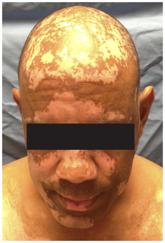
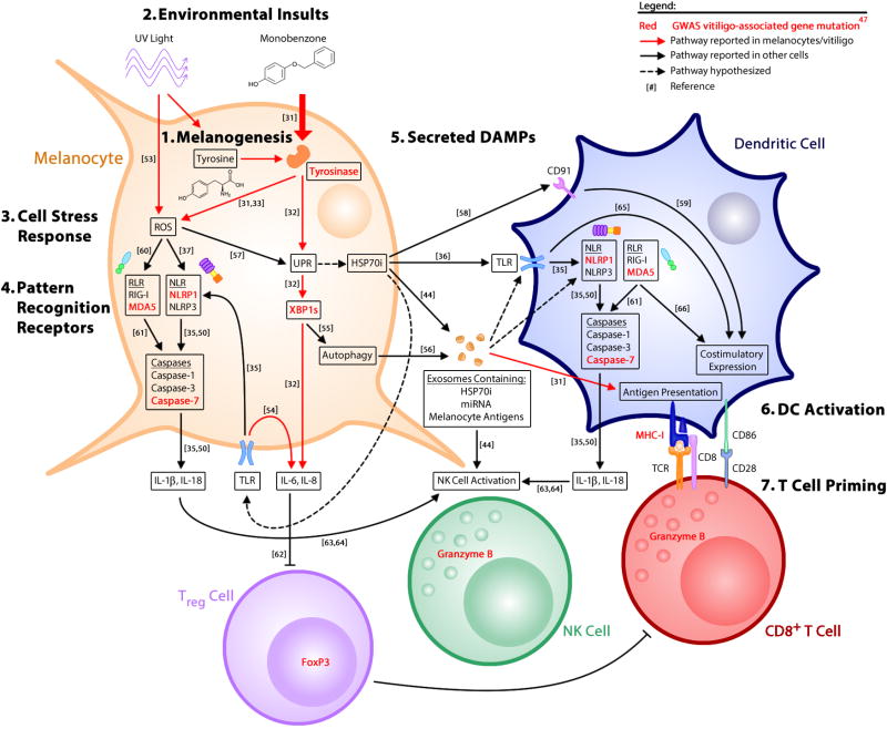
Vitiligo is an autoimmune disease of the skin in which melanocytes are destroyed by antigen-specific T cells, resulting in patchy depigmentation. Although adaptive immunity plays a clear role in disease progression, initiating factors are largely unknown. Many studies report that cellular stress pathways are dysregulated in melanocytes from vitiligo patients, suggesting that melanocyte-intrinsic defects participate in disease pathogenesis. Recent studies reveal that melanocyte stress generates damage-associated molecular patterns that activate innate immunity, thus connecting stress to organ-specific inflammation. Genetic studies in vitiligo support a role for stress, innate immunity, and adaptive mechanisms. Here, we discuss advances in the field that highlight how cellular stress, endogenous danger signals, and innate immune activation promote the onset of vitiligo.
Copyright © 2013 Elsevier Ltd. All rights reserved.
Figures
References
-
- Ongenae K, Beelaert L, van Geel N, Naeyaert JM. Psychosocial effects of vitiligo. J Eur Acad Dermatol Venereol. 2006;20:1–8. - PubMed
-
- Alikhan A, Felsten LM, Daly M, Petronic-Rosic V. Vitiligo: a comprehensive overview Part I. Introduction, epidemiology, quality of life, diagnosis, differential diagnosis, associations, histopathology, etiology, and work-up. J Am Acad Dermatol. 2011;65:473–491. - PubMed
-
- Schallreuter KU, Bahadoran P, Picardo M, Slominski A, Elassiuty YE, Kemp EH, Giachino C, Liu JB, Luiten RM, Lambe T, et al. Vitiligo pathogenesis: autoimmune disease, genetic defect, excessive reactive oxygen species, calcium imbalance, or what else? Exp Dermatol. 2008;17:139–140. discussion 141–160. - PubMed
Publication types
MeSH terms
Grants and funding
LinkOut - more resources
Full Text Sources
Other Literature Sources
Medical
