. 2013 Nov 27;5(4):986-96.
doi: 10.1016/j.celrep.2013.10.019. Epub 2013 Nov 14.

Hijacking multivesicular bodies enables long-term and exosome-mediated long-distance action of anthrax toxin

Affiliations

Hijacking multivesicular bodies enables long-term and exosome-mediated long-distance action of anthrax toxin

Laurence Abrami et al. Cell Rep. .

Abstract

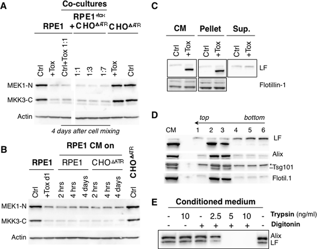
Anthrax lethal toxin is a classical AB toxin comprised of two components: protective antigen (PA) and lethal factor (LF). Here, we show that following assembly and endocytosis, PA forms a channel that translocates LF, not only into the cytosol, but also into the lumen of endosomal intraluminal vesicles (ILVs). These ILVs can fuse and release LF into the cytosol, where LF can proteolyze and disable host targets. We find that LF can persist in ILVs for days, fully sheltered from proteolytic degradation, both in vitro and in vivo. During this time, ILV-localized LF can be transmitted to daughter cells upon cell division. In addition, LF-containing ILVs can be delivered to the extracellular medium as exosomes. These can deliver LF to the cytosol of naive cells in a manner that is independent of the typical anthrax toxin receptor-mediated trafficking pathway, while being sheltered from neutralizing extracellular factors of the immune system.


Figures

Figure 1
Figure 1. LF is degraded in the cytosol by the proteasome in a translocation dependent manner
RPE1 cells were pretreated (BDEF) or not (AC) with 10 µM MG132 for 45 min or treated with 250 µg/ml Leupeptin (Leu) for 8 hrs or left untreated (Ctrl). Cells were then incubated with PA83 WT (ABE) or F427A (CDF) and LF and incubation with toxin-free medium for different time points. PNSs (40 µg) were analyzed by SDS-PAGE and Western blotting for LF, PA monomer (PAmono), PA SDS-resistant-oligomer (PAoligo) and the N-terminus of MEK1 (MEK1-N). BD. LF levels were quantified using the Typhoon or Fusion Imager and normalized to the level of LF at 1 h post toxin treatment. Error bars represent the standard deviation.
Figure 2
Figure 2. LF degradation is governed by its translocation into intraluminal vesicles
AB. RPE1 cells were transfected with siRNA against SNX3, or VSVG as a control (Ctrl siRNA). On day 3 post-transfection, cells were pretreated with 10 µM MG132 for 45 min or 250 µg/ml Leupeptin (Leupep.) for 8 h or were left untreated (Ctrl). Cells were treated with LT, followed by incubation in toxin-free medium for different time points. PNSs (40 µg) were analyzed by SDS-PAGE and Western blotting for LF. LF levels were quantified using the Fusion Imager and normalized to the level of LF at 1hr. Error bars represent the standard deviation. C–E: Schematic representation of anthrax LF in endosomes when escorted by PAWT (C) or PAF427A (E) under control conditions or by PAWT in SNX3 siRNA treated cells (D). The PA oligomer is represented in yellow for PAWT and in red for PAF427A. LF is shown in orange. The proteasome in light blue (sensitive to MG132), lysosomal enzymes in dark blue (sensitive to Leupeptin).
Figure 3
Figure 3. Exposure to anthrax LT leads to prolonged MAPKK cleavage
A. RPE1 cells were incubated with or without LT and incubation in toxin-free medium for various amounts of time. PNS (40 µg) were analyzed by SDS-PAGE and Western blotting for MEK1 N-terminus (MEK1-N), MKK3 C-terminus (MKK3-C), LF, PA monomer (PAmono), PA SDS-resistant-oligomer (PAoligo), and tubulin (as an equal-loading control). B. Western blots showing cleavage of MEK1 and MEK 2 N-terminus at various times post PA + LF treatment (45 ug each, IV) in mice. The * indicates a cross-reactive band indicative of equal loading. C. Control RPE1 cells were treated with or without LT for 1hr, and then incubated in toxin free medium for various times. A second set of RPE1 cells were pretreated 2 hrs before toxin addition with 100 nM Bafilomycin A (Baf A). After toxin treatment, these cells were incubated at room temperature for 5 min with acid buffer at pH 4.5 to allow toxin entry through the plasma membrane (acid) in the presence of Bafilomycin. PNSs (40 µg) were analyzed by SDS-PAGE and Western blotting with antibodies against LF and the MEK1 N-terminus, Actin level was used to assess equal loading. Levels of MEK-1 N-terminal reactive bands were quantified using the Typhoon scanner and normalized to 100% with non-treated cells. Error bars represent the standard deviation. D. RPE1 cells were transfected with siRNA against SNX3, or VSVG as a control (−) for 3 days before the of LT or medium, followed by washes and incubation in toxin-free medium for several hours. The isolated PNSs (40 µg) were analyzed by SDS-PAGE and Western blotting for the MEK-1 N-terminus (MEK1-N), MKK3 C-terminus (MKK3-C), and tubulin (loading control) levels.
Figure 4
Figure 4. Alteration of the MAPKK recovery kinetics upon interference with ILV dynamics
A. Schematic representation of the timeline of the experiments aimed at affecting ILV dynamics. B. RPE1 cells were incubated or not (Ctrl) with LT, followed by toxin-free medium for 4 days. At day 4, 100nM of Bafilomycin A was added to the cells in complete medium, and cell extracts were prepared at the indicated time points. PNS extracted from the samples (40 µg) were analyzed by SDS-PAGE and Western blotting against MEK1-N, MKK3-C, and tubulin. This is a representative experiment out of 3 similar studies. C. RPE1 cells were incubated with or without LT for 1hr, followed by toxin-free medium for 4 days. On day 4, samples were collected, U18666A (U18.) or anti-LBPA was added to the cells in complete medium for 24 hrs. On day 5, samples were collected, or cells that had been treated with U18666A were washed for 24 hrs and collected at day 6 (U18. w/o). PNS extracted from the samples (40 µg) were analyzed by SDS-PAGE and Western blotting against MEK1-N, MKK3-C, and tubulin. A representative experiment of three is shown. D. RPE1 cells were with or without LT for 1hr and then incubated in toxin-free medium. On day 2, cells were transfected with siRNA against TSG101, SNX3, Alix, p14, or VSVG as a control (Ctrl), or left untreated. Note that as opposed to in Figs. 2& 3, SNX3 was silenced 2 days after toxin treatment here. On day 3, a second set of toxin-treated cells were transfected with SAR1a-GTP cDNA or VPS4 DN cDNA (d5). On day 5, PNSs were extracted and analyzed by SDS-PAGE (40 µg of protein per lane) followed by Western blotting against MEK1-N, MKK3-C, TSG101, Alix, VPS4 DN –GFP, and tubulin as an equal loading control. PNS from VPS4 DN-expressing cells were also extracted on day 7 and analyzed as above. This is a representative experiment out of 3 with similar findings.
Figure 5
Figure 5. LF is present in extracellular medium of toxin treated cells
A. On day 0, RPE1 cells or CHO ΔATR (which lacks anthrax toxin receptors) were incubated with LT or medium and then in toxin-free medium for 1 day. On day 1, RPE1 and CHO ΔATR were mixed at the indicated ratio. On day 4, PNSs (40 µg) were analyzed by SDS-PAGE and Western blotting for MEK1-N, MKK3-C and actin. B. On day 0, RPE1 cells were with LT, followed by washes and toxin-free medium for 1 day. On day 1, the Conditioned Medium (CM) from RPE1 treated cells was collected and added to non-treated RPE1 cells or non-treated CHO ΔATR cells for different times as indicated. PNSs (40 µg) were analyzed by SDS-PAGE and Western blotting against MEK1-N, MKK3-C and actin. C. RPE1 cells were incubated with LT, followed by toxin-free medium for 1 day. After 1 day, the Conditioned Medium (CM) from RPE1 treated cells was collected and ultra-centrifuged for 1 hr at 100 000 × g at 4°C. The pellet and the acetone-precipitated supernatant (sup.) were dissolved in sample buffer and were analyzed and compared with 1/10th of the CM by SDS-PAGE and Western blotting against LF and Flotillin 1. D. RPE1 cells were incubated with LT, followed by toxin-free medium for 1 day. After 1 day, the Conditioned Medium (CM) from RPE1 treated cells was collected and loaded on the bottom of a sucrose step gradient (30/40/15 % sucrose). After 2 hrs of ultra-centrifugation at 100 000 × g at 4°C, 6 fractions were collected from the top, precipitated, and analyzed by SDS-PAGE and Western blotting for LF, Alix, TSG101, and Flotillin 1. E. RPE1 cells were incubated with LT, followed by toxin-free medium for 1 day. After 1 day, the CM from RPE1 treated cells was collected, treated with trypsin and/or digitonin for 30 min at room temperature. Samples were analyzed by SDS-PAGE and Western blotting performed for Alix and LF. All experiments in this figure are representative of at least 3 experiments.
Figure 6
Figure 6. LF is present in exosomes
A. RPE1 cells were transfected or not with siRNA against TSG101, SNX3, Alix, RAB11, RAB27a, RAB35, or VSVG as a control (Ctrl). After 3 days, RPE1 cells were incubated or not (Ctrl) with LT, followed by toxin-free medium for 4 hr or 1 day. After 1 day, CM from RPE1 treated cells was collected, and added to naïve RPE1 cells. For each condition, PNS (40 µg) was analyzed by SDS-PAGE and Western blotting against MEK1-N, MKK3-C and actin. B. The level of LF and Flotillin 1 was analyzed by SDS-PAGE and Western blotting in the CM described above in A. C. Naïve RPE1 cells were transfected with siRNA against TSG101, Alix, Dynamins 1 and 2, Rab27a, Rab27b, Rab35, Rab11 or VSVG as a control (Ctrl). After 2 days of transfection, these cells CM. One day later, PNSs (40 µg) were analyzed by SDS-PAGE and Western blotting against MEK1-N, MKK3-C, and actin. All experiments in this figure are representative out of at least 3 experiments.
Figure 7
Figure 7. Schematic representation of the long-term and long-distance LF delivery modes
Anthrax LT is internalized via a dynamin dependent pathway and delivered to early endosomes. There the toxin receptor complex is sorted into nascent ILVs. Anthrax PA forms a channel into the membrane of the ILV and translocates LF into the lumen. LF, encapsulated in the ILVs is transported in a microtubule dependent manner to later stages of the endocytic pathway. There, two fates are observed: LF is either delivered to the cytosol, presumably by back fusion, over periods of days or LF-containing ILVs are released into the extracellular medium as exosomes. These exosomes can be taken up by naïve recipient cell via an anthrax toxin receptor independent dynamin-dependent mechanism. LF is subsequently released into the cytosol of the recipient cell in a Tgs101 and Alix dependent manner (inspired from (Raposo and Stoorvogel, 2013)).

References

    1. Abrami L, Bischofberger M, Kunz B, Groux R, van der Goot FG. Endocytosis of the anthrax toxin is mediated by clathrin, actin and unconventional adaptors. PLoS Pathogens. 2010;6:e1000792. - PMC - PubMed
    1. Abrami L, Lindsay M, Parton RG, Leppla SH, van der Goot FG. Membrane insertion of anthrax protective antigen and cytoplasmic delivery of lethal factor occur at different stages of the endocytic pathway. J Cell Biol. 2004;166:645–651. - PMC - PubMed
    1. Alouf JE, Freer JH, editors. The comprehensive sourcebook of bacterial protein toxins 3rd Edition. London: Academic Press; 2005.
    1. Baietti MF, Zhang Z, Mortier E, Melchior A, Degeest G, Geeraerts A, Ivarsson Y, Depoortere F, Coomans C, Vermeiren E, et al. Syndecan-syntenin-ALIX regulates the biogenesis of exosomes. Nature cell biology. 2012;14:677–685. - PubMed
    1. Baldari CT, Tonello F, Paccani SR, Montecucco C. Anthrax toxins: A paradigm of bacterial immune suppression. Trends Immunol. 2006;27:434–440. - PubMed

Publication types