

In vivo thermal ablation monitoring using ultrasound echo decorrelation imaging
- PMID: 24239361
- PMCID: PMC3849110
- DOI: 10.1016/j.ultrasmedbio.2013.09.007
In vivo thermal ablation monitoring using ultrasound echo decorrelation imaging
Abstract
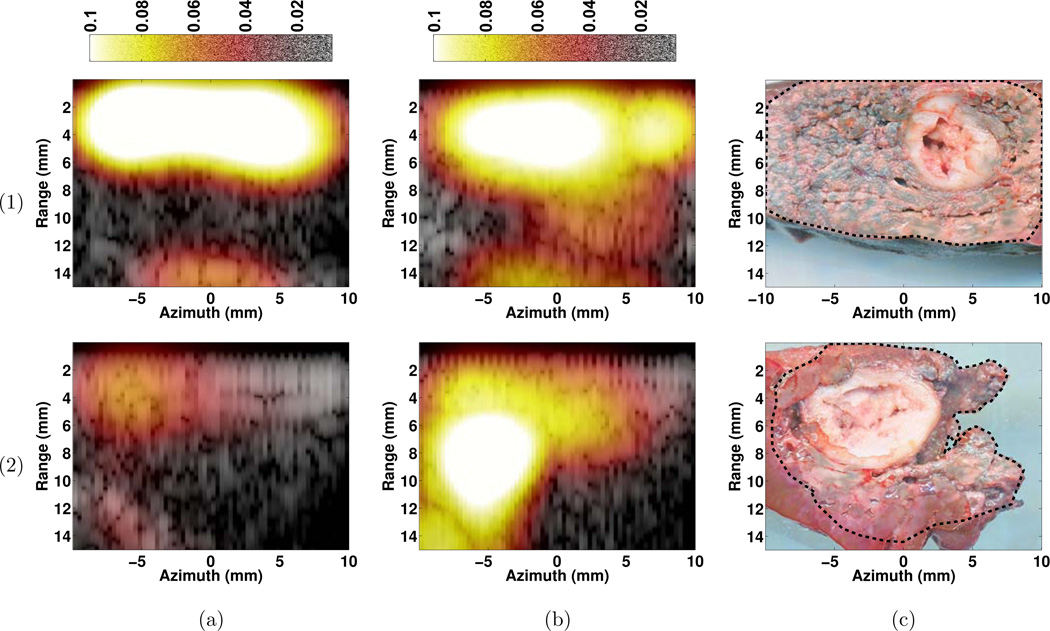
Previous work indicated that ultrasound echo decorrelation imaging can track and quantify changes in echo signals to predict thermal damage during in vitro radiofrequency ablation (RFA). In the in vivo studies reported here, the feasibility of using echo decorrelation imaging as a treatment monitoring tool was assessed. RFA was performed on normal swine liver (N = 5), and ultrasound ablation using image-ablate arrays was performed on rabbit liver implanted with VX2 tumors (N = 2). Echo decorrelation and integrated backscatter were computed from Hilbert transformed pulse-echo data acquired during RFA and ultrasound ablation treatments. Receiver operating characteristic (ROC) curves were employed to assess the ability of echo decorrelation imaging and integrated backscatter to predict ablation. Area under the ROC curves (AUROC) was determined for RFA and ultrasound ablation using echo decorrelation imaging. Ablation was predicted more accurately using echo decorrelation imaging (AUROC = 0.832 and 0.776 for RFA and ultrasound ablation, respectively) than using integrated backscatter (AUROC = 0.734 and 0.494).
Keywords: Bulk ultrasound ablation; Echo decorrelation imaging; Radiofrequency ablation; Therapy monitoring.
Copyright © 2014 World Federation for Ultrasound in Medicine & Biology. Published by Elsevier Inc. All rights reserved.
Figures


References
-
- Arora D, Cooley D, Perry T, Guo J, Parker D, Skliar M, Roemer R. Thermal dose control of ultrasound therapies using MR thermometry images: an in-vitro phantom study. In: IEEE American Control Conference 2005. IEEE. 2005:405–410.
-
- Arthur RM, Straube WL, Trobaugh JW, Moros EG. Non-invasive estimation of hyperthermia temperatures with ultrasound. Int J Hyperthermia. 2005;216:589–600. - PubMed
-
- Barthe P, Slayton M, Jaeger P, Makin I, Gallagher L, Mast T, Runk M, Faidi W. Ultrasound therapy system and ablation results utilizing miniature imaging/therapy arrays. IEEE Ultrasonics Symposium. 2004;Vol. 3:1792–1795. Vol.3.
-
- Boaz T, Lewin J, Chung Y, Duerk J, Clampitt M, Haaga J. MR monitoring of MR-guided radiofrequency thermal ablation of normal liver in an animal model. J Magn Reson Imaging. 1998;81:64–69. - PubMed
-
- Caspani B, Ierardi AM, Motta F, Cecconi P, Fesce E, Belli L. Small nodular hepatocellular carcinoma treated by laser thermal ablation in high risk locations: preliminary results. Eur Radiol. 2010;209:2286–2292. - PubMed
Publication types
MeSH terms
Grants and funding
LinkOut - more resources
Full Text Sources
Other Literature Sources
Medical
