. 2014 Jan 7;22(1):104-15.
doi: 10.1016/j.str.2013.10.001. Epub 2013 Nov 14.

Alternate splicing of dysferlin C2A confers Ca²⁺-dependent and Ca²⁺-independent binding for membrane repair

Affiliations

Alternate splicing of dysferlin C2A confers Ca²⁺-dependent and Ca²⁺-independent binding for membrane repair

Kerry Fuson et al. Structure. .

Abstract

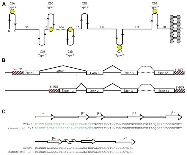
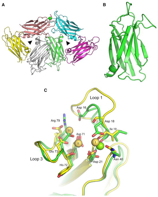
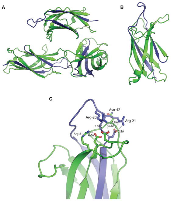
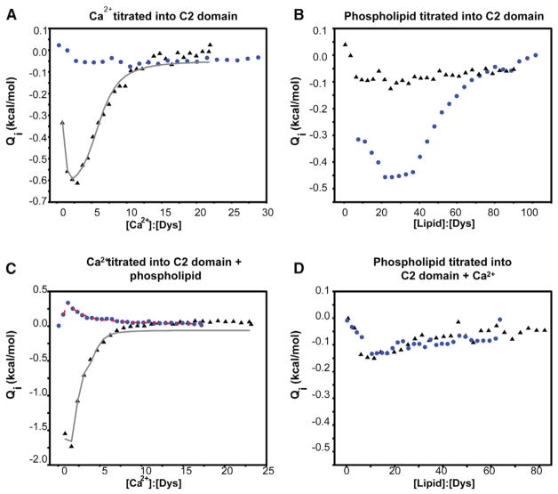
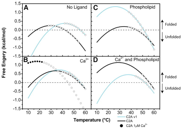
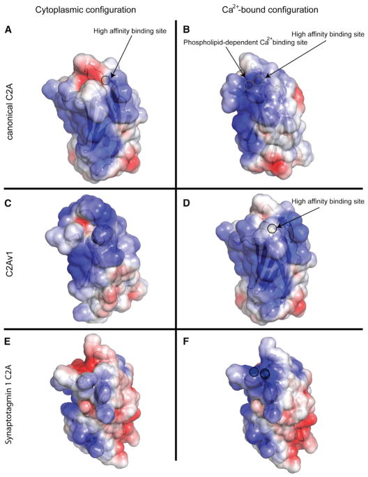
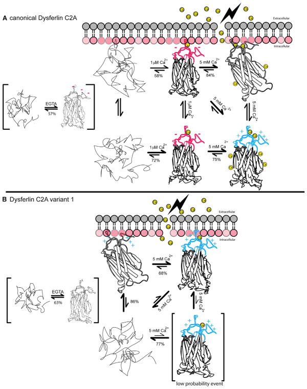
Dysferlin plays a critical role in the Ca²⁺-dependent repair of microlesions that occur in the muscle sarcolemma. Of the seven C2 domains in dysferlin, only C2A is reported to bind both Ca²⁺ and phospholipid, thus acting as a key sensor in membrane repair. Dysferlin C2A exists as two isoforms, the "canonical" C2A and C2A variant 1 (C2Av1). Interestingly, these isoforms have markedly different responses to Ca²⁺ and phospholipid. Structural and thermodynamic analyses are consistent with the canonical C2A domain as a Ca²⁺-dependent, phospholipid-binding domain, whereas C2Av1 would likely be Ca²⁺-independent under physiological conditions. Additionally, both isoforms display remarkably low free energies of stability, indicative of a highly flexible structure. The inverted ligand preference and flexibility for both C2A isoforms suggest the capability for both constitutive and Ca²⁺-regulated effector interactions, an activity that would be essential in its role as a mediator of membrane repair.


Figures

Figure 1
Figure 1. Overall Schematic of Dysferlin Domain Structure, Exon Structure, and C2A Primary/Secondary Sequence
(A) Overall modular organization of the full-length dysferlin protein. The spacers between C2 domains have been scaled according to the predicted distances between C2 domains. Yellow spheres correspond to predicted Ca2+-binding C2 domains in dysferlin based on conserved residues in the primary sequence alignments. The dotted red line corresponds to the FerA/B and DysN/DysC domains. (B) Schematic of the partial exon structure of dysferlin C2A. (C) Primary sequence alignment of C2A variant 1 versus the canonical C2A domain of dysferlin. The arrows above the sequence correspond to residues that possess β strand secondary structure, whereas the helical cartoon corresponds to residues with α-helical secondary structure.
Figure 2
Figure 2. Crystal Structure of the Canonical Dysferlin C2A Domain
(A) Ribbon diagram of the stacked trimeric arrangement of the asymmetric unit of human dysferlin C2A. The green sphere is a single Ca2+ coordinated to one of the six domains in the asymmetric unit. Noncrystallographic 3-fold symmetry operators are represented as triangles. (B) An isolated canonical C2A domain is shown in green. (C) Ca2+ binding sites in dysferlin C2A. The green sticks correspond to the dysferlin C2A structure. The green sphere corresponds to the high-affinity Ca2+ found in the crystal structure (chain E, 4IHB). The yellow sticks correspond to the three La3+ described in the phospholipase C-δ1 C2 domain (1DJG, residues 628–756) (Essen et al., 1997). The residue numbers refer to the corresponding amino acids in the human dysferlin sequence.
Figure 3
Figure 3. Crystal Structure of the Human Dysferlin C2A Variant 1 Domain
(A) Asymmetric unit of C2Av1. (B) Single C2Av1 domain. (C) Cation binding pocket of dysferlin C2Av1. The variant residues are shown in blue.
Figure 4
Figure 4. Ligand Binding Profiles for the Canonical Dysferlin C2A and the C2Av1 Domains
The canonical construct is shown above as black triangles, whereas the C2Av1 construct is shown as blue circles. The fits of the canonical C2A data are shown as a gray line, whereas the fits of the C2Av1 data are shown as a dashed red line. All heats were normalized to the concentration of protein in the sample cell at each injection. (A) Titration of 102 μM dysferlin C2A and the titration of 103 μM C2Av1 with Ca2+. (B) Titration of 50 μM dysferlin C2A and the titration of 56 μM C2Av1 with LUVs made of a 60:40 mixture of POPC:POPS. (C) Titration of 80 μM dysferlin C2A and the titration of 108 μM dysferlin C2Av1 with Ca2+ in the presence of 5 mM total lipid composed of LUVs made of a 60:40 mixture of POPC:POPS. (D) Titration of 47 μM dysferlin C2A and the titration of 90 μM dysferlin C2Av1 with LUVs made of a 60:40 mixture of POPC:POPS in the presence of 2 mM Ca2+.
Figure 5
Figure 5. Free Energy Diagrams of Dysferlin C2A Domains
In the absence of Ca2+ (A), Ca2+ (B), phospholipid (C), and phospholipid and Ca2+ (D). At any point along the curve, the native and denatured states of the C2A domains exist at varying ratios. As the temperature changes so does the ratio between the two states. Where the curve crosses 0 kcal/mol on the y axis, the populations of protein in the native and denatured states are equal to one another; below this, the protein is found predominantly in the denatured state, and above this, the protein is predominantly found in the native state. The open circles on each curve represent the temperature over which the protein denatured.
Figure 6
Figure 6. Electrostatic Surface Potential of Dysferlin C2 Domains
(A and B) The canonical C2A domain with (A) a single Ca2+ and canonical C2A with two Ca2+ (B). (C and D) Dysferlin C2Av1 with no ligand (C) and a single Ca2+ (D). Solvent-accessible surface is colored by the calculated electrostatic potential and displayed at ± 3 kT/e. Calcium ions are highlighted by a circular outline. (E and F) The electrostatic surface potential representation of synaptotagmin 1 C2A with and without (E) and with (F) saturating Ca2+ (1byn) is shown for comparison (Shao et al., 1998). Calcium ions are highlighted by a circular outline.
Figure 7
Figure 7. Full-Length Dysferlin Proteins Bearing Alternately Spliced Canonical C2A or C2Av1 Show Similar Intrinsic Capacity to Target the Plasma Membrane
(A) Representative confocal images showing surface-expressed EGFP-FLC2ADysferlinMycHis or EGFP-FLC2Av1DysferlinMycHis. Transfected C2C12 myoblasts were incubated with mouse anti-His to selectively label the extracellular His epitope. Labeling was performed on live cells at <10°C to prevent endosomal internalization of surface-bound antibody. Cells were then washed, fixed, and labeled with goat anti-mouse555. Scale bar 10 μM. (B) Flow cytometry quantifies similar levels of surface-expressed EGFP-FLC2ADysferlinMycHis or EGFP-FLC2Av1DysferlinMycHis. Transfected C2C12 myoblasts were dissociated from the culture dish and labeled as live cells at <10°C with mouse anti-His followed by an anti-mousealexa647 secondary antibody. Live cells were gated based on impermeability to propidium iodide (data not shown). (Left panel) Shows increasing levels of surface-labeled anti-Hisalexa647 (x axis) is proportional to the levels of EGFP auto-fluorescence (y axis). Gates for transfected (Tfd) and untransfected (Un) cells are shown; very highly transfected cells often show signs of toxicity and were excluded from analysis. (Middle panel) Histogram showing similar normal distributions of surface bound anti-mousealexa647 in populations of transfected cells expressing FLC2A or FLC2Av1 constructs, from duplicate samples labeled on the same day. (Right panel) Pooled data from three experiments performed in duplicate showing similar levels of surface-labeled anti-Hisalexa647 relative to EGFP autofluorescence for both FLC2A or FLC2Av1 constructs. To allow comparison between constructs transfected and labeled on the same day, values derived from canonical C2A were normalized to one. Error bars are reported as the 95% confidence interval.
Figure 8
Figure 8. Summary of Thermodynamic and Structural Results
(A) Thermodynamic interactions among the canonical C2A domain of dysferlin, Ca2+, and the negatively charged phospholipid (red lollipop representation of a lipid surface). Unfolded protein is shown as a random trace. The various folded states of the C2A domain are shown as a partially unfolded domain with intact Ca2+:phospholipid binding loops at the top of the structure. Calcium ions are shown as yellow spheres. The lightning bolt on the extracellular side of the membrane represents the site of membrane damage. Red color designates net negative charge; blue color designates net positive charge. The relative equilibrium of the various measure states is also shown. Percentages underneath the equilibrium arrows are the % folded protein under the stated equilibrium conditions. The structures within brackets likely do not exist in the muscle cell, but the thermodynamic relationship was described. (B) Thermodynamic interactions among the C2A variant, Ca2+, and negatively charged phospholipid. The single structure within brackets (C2Av1 + Ca2+) is a low probability event.

Similar articles

Cited by

References

    1. Ampong BN, Imamura M, Matsumiya T, Yoshida M, Takeda S. Intracellular localization of dysferlin and its association with the dihydropyridine receptor. Acta Myol. 2005;24:134–144. - PubMed
    1. Anandakrishnan R, Aguilar B, Onufriev AV. H++ 3.0: automating pK prediction and the preparation of biomolecular structures for atomistic molecular modeling and simulations. Nucleic Acids Res. 2012;40:W537–W541. Web Server issue. - PMC - PubMed
    1. Azakir BA, Di Fulvio S, Therrien C, Sinnreich M. Dysferlin interacts with tubulin and microtubules in mouse skeletal muscle. PLoS ONE. 2010;5:e10122. - PMC - PubMed
    1. Bansal D, Miyake K, Vogel SS, Groh S, Chen CC, Williamson R, McNeil PL, Campbell KP. Defective membrane repair in dysferlin-deficient muscular dystrophy. Nature. 2003;423:168–172. - PubMed
    1. Battye TG, Kontogiannis L, Johnson O, Powell HR, Leslie AG. iMOSFLM: a new graphical interface for diffraction-image processing with MOSFLM. Acta Crystallogr D Biol Crystallogr. 2011;67:271–281. - PMC - PubMed

Publication types

MeSH terms