Review
. 2014 Jan 7;19(1):8-20.
doi: 10.1016/j.cmet.2013.10.003. Epub 2013 Nov 14.

Weighing in on adipocyte precursors

Affiliations
Review

Weighing in on adipocyte precursors

Ryan Berry et al. Cell Metab. .

Abstract

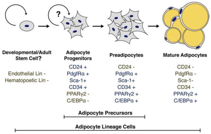
Obesity, defined as an excessive increase in white adipose tissue (WAT), is a global health epidemic. In obesity, WAT expands by increased adipocyte size (hypertrophy) and number (hyperplasia). The location and cellular mechanisms of WAT expansion greatly affect the pathogenesis of obesity. However, the cellular and molecular mechanisms regulating adipocyte size, number, and depot-dependent expansion in vivo remain largely unknown. This perspective summarizes previous work addressing adipocyte number in development and obesity and discusses recent advances in the methodologies, genetic tools, and characterization of in vivo adipocyte precursor cells allowing for directed study of hyperplastic WAT growth in vivo.


Figures

Figure 1
Figure 1. Identification of mature adipocyte nuclei in situ
Images of whole mounted white adipose tissue. The lipid is stained with LipidTox (green), the plasma membrane with Cell Mask Orange (red), and the nuclei with DAPI (blue). Most nuclei in WAT are in close proximity to lipid droplets. Therefore accurate identification of adipocyte nuclei (white arrowheads) from non-adipocyte nuclei (yellow arrowheads) requires staining of the plasma membrane. Nuclei that are not separated from the lipid droplets by a plasma membrane are the mature adipocyte nuclei. Scale bars = 25μm.
Figure 2
Figure 2. A model of in vivo adipogenesis
A schematic representation of the adipocyte cellular lineage is shown. The defined lineage, cell surface, and transcriptional markers for each cell type are shown below each listed cell type. Positive markers are shown in blue while negative markers are shown in gold. The question marks highlight that it is currently unclear if the adipocyte progenitor population contains self-renewing adipocyte stem cells or if the adipocyte progenitors are repopulated by another, unidentified population of adipocyte stem cells in adults.
Figure 3
Figure 3. Adipocyte progenitors in WAT depots
CD24+ adipocyte progenitor cells are a distinct population of WAT resident stromal cells. Shown are dot plots of the Lin-:CD29+:CD34+ cell populations from the indicated depots. The cells are present in levels as high at 2% in adult SWAT depots. Cells with an identical cell surface marker profile are present in VWAT depots, but are less abundant (peri-gonadal WAT (GWAT) is shown).

References

    1. Anderson DB, Kauffman RG. Cellular and enzymatic changes in porcine adipose tissue during growth. J Lipid Res. 1973;14:160–168. - PubMed
    1. Berg W. The development of human fat. Z. Morph. Anthrop. 1911;13
    1. Bernlohr DA, Bolanowski MA, Kelly TJ, Lane MD. Evidence for an increase in transcription of specific mRNAs during differentiation of 3T3-L1 preadipocytes. J Biol Chem. 1985;260:5563–5567. - PubMed
    1. Berry R, Rodeheffer MS. Characterization of the adipocyte cellular lineage in vivo. Nat Cell Biol. 2013;15:302–308. - PMC - PubMed
    1. Billon N, Iannarelli P, Monteiro MC, Glavieux-Pardanaud C, Richardson WD, Kessaris N, Dani C, Dupin E. The generation of adipocytes by the neural crest. Development. 2007;134:2283–2292. - PMC - PubMed

Publication types