

Proteasomal and lysosomal protein degradation and heart disease
- PMID: 24239609
- PMCID: PMC4011941
- DOI: 10.1016/j.yjmcc.2013.11.006
Proteasomal and lysosomal protein degradation and heart disease
Abstract
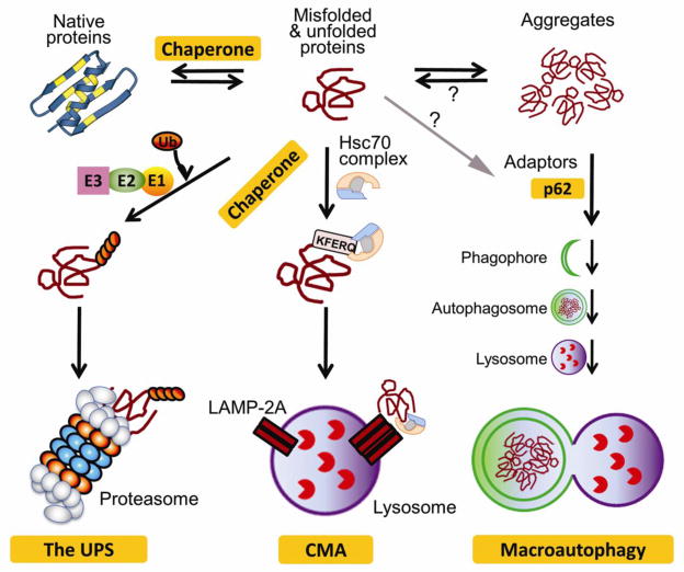
In the cell, the proteasome and lysosomes represent the most important proteolytic machineries, responsible for the protein degradation in the ubiquitin-proteasome system (UPS) and autophagy, respectively. Both the UPS and autophagy are essential to protein quality and quantity control. Alterations in cardiac proteasomal and lysosomal degradation are remarkably associated with most heart disease in humans and are implicated in the pathogenesis of congestive heart failure. Studies carried out in animal models and in cell culture have begun to establish both sufficiency and, in some cases, the necessity of proteasomal functional insufficiency or lysosomal insufficiency as a major pathogenic factor in the heart. This review article highlights some recent advances in the research into proteasome and lysosome protein degradation in relation to cardiac pathology and examines the emerging evidence for enhancing degradative capacities of the proteasome and/or lysosome as a new therapeutic strategy for heart disease. This article is part of a Special Issue entitled "Protein Quality Control, the Ubiquitin Proteasome System, and Autophagy".
Keywords: Autophagy; Heart disease; Lysosome; Proteasome; Ubiquitin.
Copyright © 2013 Elsevier Ltd. All rights reserved.
Conflict of interest statement
No conflicts of interest, financial or otherwise are declared by the authors
Figures
References
-
- Willis MS, Patterson C. Proteotoxicity and cardiac dysfunction--Alzheimer’s disease of the heart? N Engl J Med. 2013;368:455–64. - PubMed
-
- Gomes AV, Zong C, Edmondson RD, Berhane BT, Wang GW, Le S, et al. The murine cardiac 26S proteasome: an organelle awaiting exploration. Ann N Y Acad Sci. 2005;1047:197–207. - PubMed
Publication types
MeSH terms
Substances
Grants and funding
- P01 HL069779/HL/NHLBI NIH HHS/United States
- P01 HL059408/HL/NHLBI NIH HHS/United States
- R01HL072166/HL/NHLBI NIH HHS/United States
- R01 HL062927/HL/NHLBI NIH HHS/United States
- R01 HL085629/HL/NHLBI NIH HHS/United States
- R01HL085629/HL/NHLBI NIH HHS/United States
- R011062927/PHS HHS/United States
- R01HL068936/HL/NHLBI NIH HHS/United States
- R01 HL087862/HL/NHLBI NIH HHS/United States
- R01 HL072166/HL/NHLBI NIH HHS/United States
- P01HL059408/HL/NHLBI NIH HHS/United States
- P01HL69779/HL/NHLBI NIH HHS/United States
- R01HL05924/HL/NHLBI NIH HHS/United States
- R01 HL068936/HL/NHLBI NIH HHS/United States
LinkOut - more resources
Full Text Sources
Other Literature Sources
Medical
