Review
. 2013 Nov 15;27(22):2409-26.
doi: 10.1101/gad.228080.113.

Building the mammalian testis: origins, differentiation, and assembly of the component cell populations

Affiliations
Review

Building the mammalian testis: origins, differentiation, and assembly of the component cell populations

Terje Svingen et al. Genes Dev. .

Abstract

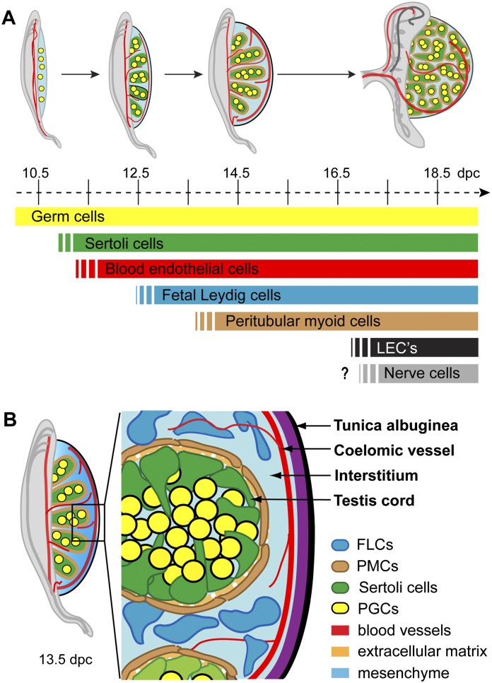
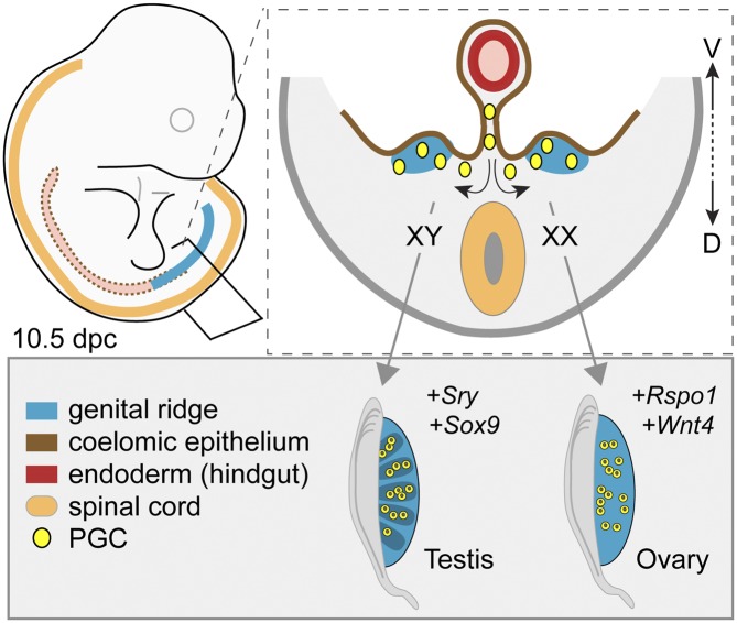
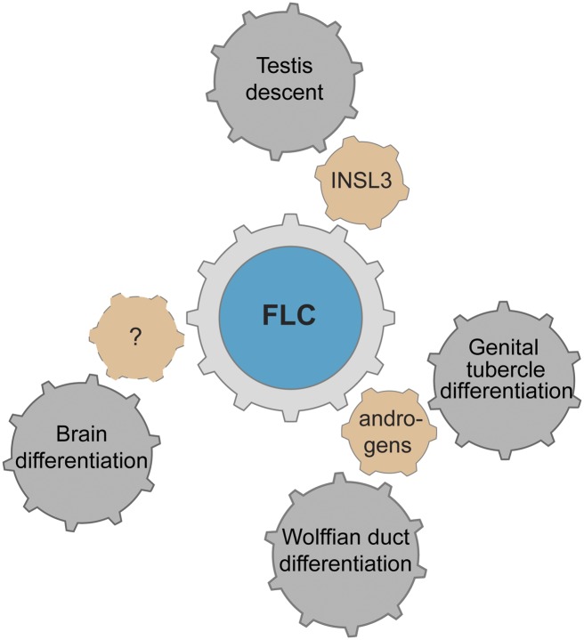
Development of testes in the mammalian embryo requires the formation and assembly of several cell types that allow these organs to achieve their roles in male reproduction and endocrine regulation. Testis development is unusual in that several cell types such as Sertoli, Leydig, and spermatogonial cells arise from bipotential precursors present in the precursor tissue, the genital ridge. These cell types do not differentiate independently but depend on signals from Sertoli cells that differentiate under the influence of transcription factors SRY and SOX9. While these steps are becoming better understood, the origins and roles of many testicular cell types and structures-including peritubular myoid cells, the tunica albuginea, the arterial and venous blood vasculature, lymphatic vessels, macrophages, and nerve cells-have remained unclear. This review synthesizes current knowledge of how the architecture of the testis unfolds and highlights the questions that remain to be explored, thus providing a roadmap for future studies that may help illuminate the causes of XY disorders of sex development, infertility, and testicular cancers.

Keywords: Leydig cells; Sertoli cells; disorder of sex development; fertility; organogenesis; sex determination.


Figures

Figure 1.
Figure 1.
Anatomy of the developing mouse testis. (A) Schedule of cell lineage differentiation during testis organogenesis. The genital ridges are colonized by germ cells (yellow) prior to testis specification. The first somatic cells to differentiate are the Sertoli cells (green), with blood endothelial cells also migrating into the gonad at this early stage to lay down a primitive arterial vasculature (red). Following a period of Sertoli cell proliferation, fetal Leydig cells (FLCs; blue) and peritubular myoid cells (PMCs; brown) differentiate. The vasculature becomes more complete with additional development of venous vessels and lymphatic endothelial cells (LECs; black). Although the postnatal testis also contains neurons, the developmental stage at which they first appear remains unclear. (B) Fetal testis architecture. By 13.5 dpc, the mouse testis is compartmentalized into testis cords and interstitial space, with the majority of the cell types of the mature testis already in place. The testis cords comprise mitotically arrested germ cells surrounded by Sertoli cells, with an outer layer of PMCs and an extracellular matrix (ECM) giving structural support. The interstitium consists of mesenchymal tissue, steroidogenic FLCs, and a prominent blood vascular network. At this stage, the protective testis cap, the tunica albuginea, has also begun to develop.
Figure 2.
Figure 2.
Gonadal sex determination: a choice between two mutually opposing fates. The genital ridges contain at least three types of unspecified, bipotential precursor cells. In XY gonads, cells of the supporting cell lineage start to express Sry and then Sox9, causing them to differentiate into Sertoli cells. In the absence of Sry, as in XX genital ridges, the same precursor cells differentiate into granulosa cells under the influence of genes encoding transcription factors, including Ctnnb1 and Foxl2. In addition to promoting the Sertoli cell differentiation pathway, Sry and Sox9 also (directly or indirectly) suppress female-specific cell differentiation pathways. Sertoli cells induce (dotted arrows) other cell populations to differentiate into the steroidogenic FLCs that otherwise would differentiate into ovarian theca cells and enter the spermatogenic pathway as opposed to the oocyte differentiation pathway.
Figure 3.
Figure 3.
The establishment of the bipotential genital ridges and gonadal sex determination. In mammals, the genital ridges (blue) typically appear as longitudinal outgrowths along the surfaces of the mesonephroi within the coelomic cavity. In mice, they emerge at ∼10 dpc through recruitment of cells from the overlying coelomic epithelium (brown). Primordial germ cells (yellow) colonize the genital ridges (arrows) after leaving the hindgut (red) via the dorsal mesentery. At this stage in development, the genital ridges are bipotential and can differentiate into testes or ovaries, depending on genetic cues. From ∼10.5 dpc, the Y-linked sex-determining gene Sry is expressed in XY genital ridges and initiates Sox9 expression and testis differentiation. In the absence of Sry, as in XX genital ridges, ovary differentiation is initiated by the action of genes such as Rspo1 and Wnt4. (D) Dorsal; (V) ventral.
Figure 4.
Figure 4.
Sertoli cells as the organizing center of testis differentiation. Sertoli cells are the first somatic cells to differentiate in the XY gonad. They then act as a regulatory hub during testis organogenesis, influencing testis cord formation, Müllerian duct regression, and the differentiation and function of several other cell types, including the germ cells, PMCs, FLCs, and endothelial cells (ECs). In turn, androgens produced by the FLCs are largely responsible for the masculinization of the embryo.
Figure 5.
Figure 5.
Known and proposed origins of the testicular cell lineages. The cells of the nascent genital ridges originate primarily from the overlying coelomic epithelium but also from the subjacent mesonephros. A subset of ingressing coelomic epithelial cells differentiates into Sertoli cells following Sry expression. Some of these supporting cells are also believed to differentiate into FLCs. It is unclear whether cells originating from the mesonephros contribute toward somatic cells other than blood endothelium, but they very likely contribute to the mesenchyme. The origin of PMCs remains unknown, but it is likely that they differentiate from a subset of mesenchymal cells or yet unidentified precursor cells of the testis interstitium. A second origin for the FLCs has also been proposed to include perivascular cells located at the gonad–mesonephric junction.
Figure 6.
Figure 6.
Correct spatiotemporal expression of Sry within the genital ridges is required for testis differentiation. (A) Sry transcription (dark blue) is initiated in a subset of supporting lineage cells in the central region of the genital ridges. From here, the domain of Sry expression extends toward the gonadal poles. In mice, Sry is expressed along the entire length of the genital ridge for only a brief period before becoming down-regulated first within the center region, then at the anterior pole, and finally at the posterior pole. (In larger mammals, including humans, Sry expression is maintained after testis differentiation.) (B,C) Experimentally, it has been shown that Sry expression must reach a threshold level within a temporal window of 14–19 ts (∼11.2–11.75 dpc; yellow shading) in order to activate Sox9 expression (green) and hence Sertoli cell differentiation and testis cord formation appropriately along the entire length of the testis. (B) Crossing Ypos mice (harboring a Sry locus that causes retarded Sry expression) with C57BL/6 mice and delaying Sry initiation by a few hours does not allow it to be expressed along the entire length of the genital ridge at the critical time, with the result that Sox9 expression (green) is limited to the central region, with the ultimate result of ovotesticular development, characterized by testicular tissue in the center and ovarian tissue at the poles. (C) By experimentally delaying Sry expression until after 11.75 dpc in transgenic mice, Sox9 is not expressed during the critical time window in the majority of supporting cells, resulting in a complete failure of Sox9 up-regulation and Sertoli cell differentiation and hence ovarian differentiation.
Figure 7.
Figure 7.
Effects of Sertoli cells on germ cell differentiation and survival. In the fetal testis, Sertoli cells influence XY germ cells via multiple signaling pathways. Degradation of retinoic acid (RA) by CYP26B1 expression together with FGF9 secretion allow gonocytes to avoid meiosis. FGF9 also stimulates expression of Cripto, which, together with Nodal, promotes maintenance of pluripotency. Factors inducing mitotic arrest are hypothesized to be produced by Sertoli cells and may include Activin A and TGFβ. Finally, several factors have been shown to influence the health and survival of germ cells postnatally, including PDGF, TGFβ, and estrogen (see the text for further details).
Figure 8.
Figure 8.
Proposed models of FLC versus ALC origins. In most mammals, the FLC population appears to perish shortly after birth, to be replaced by a new ALC population. However, different hypotheses have been proposed to explain the origins of the two populations. (A) The most popular hypothesis: The FLCs and ALCs arise from two unique precursor cell populations unrelated to each other, with the FLCs disappearing completely (cross). (B) The least-regarded hypothesis: FLCs and ALCs are the same cell lineage; the FLC population almost disappears postnatally, but a few cells remain to divide and differentiate into ALCs. (C) Alternative hypothesis gaining momentum: FLCs diminish postnatally (cross), and the ALC population is established from the same precursor cells that gave rise to FLCs after lying dormant throughout prepubertal development.
Figure 9.
Figure 9.
Factors produced by FLCs affecting male sexual development. The Leydig cells are steroidogenic cells, and both the FLCs and ALCs are responsible for producing androgens. During fetal life, androgens induce the Wolffian ducts to form epididymides, vasa deferentia, and seminal vesicles. Androgens further stimulate the elongation of the genital tubercle to form the penis and tissue fusion to form the penile shaft and scrotum. FLCs also produce factors necessary for testicular descent, including INSL3. Evidence strongly suggests that sex hormones influence differences in brain development between the sexes, but the factors involved remain unknown.

References

    1. Albrecht KH, Eicher EM 2001. Evidence that Sry is expressed in pre-Sertoli cells and Sertoli and granulosa cells have a common precursor. Dev Biol 240: 92–107 - PubMed
    1. Allen LG, Wilson FJ, Macdonald GJ 1989. Neuropeptide Y-containing nerves in the rat gonads: Sex difference and development. Biol Reprod 40: 371–378 - PubMed
    1. Anderson R, Copeland TK, Schöler H, Heasman J, Wylie C 2000. The onset of germ cell migration in the mouse embryo. Mech Dev 91: 61–68 - PubMed
    1. Anesetti G, Lombide P, D'Albora H, Ojeda SR 2001. Intrinsic neurons in the human ovary. Cell Tissue Res 306: 231–237 - PubMed
    1. Azhar S, Leers-Sucheta S, Reaven E 2003. Cholesterol uptake in adrenal and gonadal tissues: The SR-BI and ‘selective’ pathway connection. Front Biosci 8: s998–s1029 - PubMed

Publication types

LinkOut - more resources