. 2014 Jan;32(1):76-83.
doi: 10.1038/nbt.2747. Epub 2013 Nov 17.

Transient cytokine treatment induces acinar cell reprogramming and regenerates functional beta cell mass in diabetic mice

Affiliations

Transient cytokine treatment induces acinar cell reprogramming and regenerates functional beta cell mass in diabetic mice

Luc Baeyens et al. Nat Biotechnol. 2014 Jan.

Retraction in

Abstract

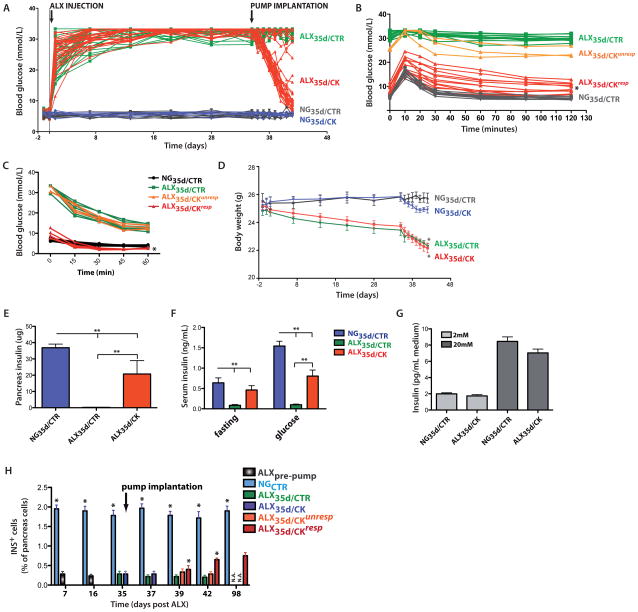
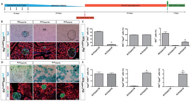
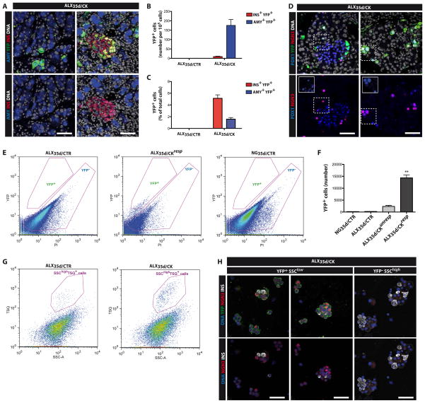
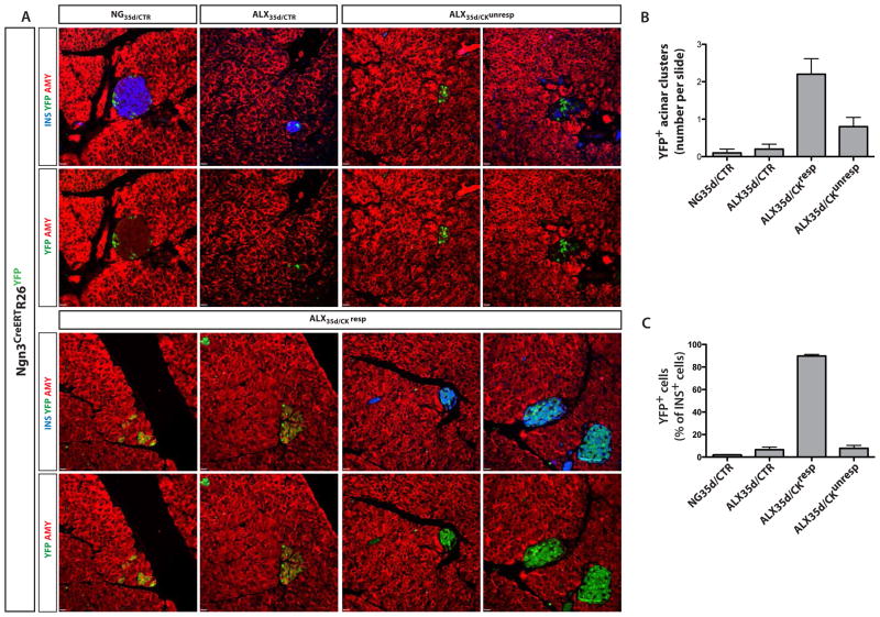
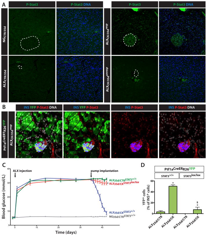
Reprogramming of pancreatic exocrine cells into cells resembling beta cells may provide a strategy for treating diabetes. Here we show that transient administration of epidermal growth factor and ciliary neurotrophic factor to adult mice with chronic hyperglycemia efficiently stimulates the conversion of terminally differentiated acinar cells to beta-like cells. Newly generated beta-like cells are epigenetically reprogrammed, functional and glucose responsive, and they reinstate normal glycemic control for up to 248 d. The regenerative process depends on Stat3 signaling and requires a threshold number of Neurogenin 3 (Ngn3)-expressing acinar cells. In contrast to previous work demonstrating in vivo conversion of acinar cells to beta-like cells by viral delivery of exogenous transcription factors, our approach achieves acinar-to-beta-cell reprogramming through transient cytokine exposure rather than genetic modification.


Figures

Figure 1
Figure 1. Chronic diabetes in mice can be reverted by transient release of EGF and CNTF
(A) Hyperglycemia was induced by intravenous (i.v.) injection of a single dose of the beta cell toxin alloxan (ALX) in 8–13 week old male mice of mixed background. Glycemia was then assayed by a portable glucometer in blood collection from the tail vein on a daily or weekly basis. Where indicated, 35 days after ALX injection, osmotic mini-pumps loaded with EGF and CNTF (ALX35d/CK, n=35) or vehicle (ALX35d/CTR, n=35) were implanted intraperitoneally (i.p.). NG35d/CTR (n=10) and NG35d/CK (n=10) mice were not treated with ALX and were implanted with vehicle and cytokine-loaded pumps, respectively. (B) I.p. glucose tolerance test (IPGTT). 2g glucose per kg BW was injected and clearance in blood was measured at indicated time points to indirectly measure the glucose responsiveness of insulin secretion by beta cells (*: p<0.001, ALX35d/CK vs ALX35d/CTR mice; n=15 each). (C) Insulin Tolerance Test (ITT). Blood glucose levels following i.v. injection of 0.75U insulin per kg BW were measured. (*p<0.01, ALX35d/CKresp (n=5) vs. NG35d/CTR mice (n=9)). (D) Body weight was measured at indicated time points. Differences in body weight between ALX-treated and untreated mice were not statistically significant until implantation of the pump on day 35 (p>0.05 from day 0 to day 34, p<0.05 from day 35 to day 42, ALX35d vs NG35d mice). (E) Total pancreas insulin content was measured by radio-immuno assay on tissue extracts at 7 days after pump implantation. (F) Serum insulin was measured 7 days after pump implantation, at time point 0 (after 6h fasting) and 30 minutes after i.p. injection of 2g glucose per kg BW (glucose) (**: p<0.01). (G) Insulin secretion by islets isolated from NG35d/CTR and ALX35d/CK mice (7 days after pump implantation). Islets were stimulated with 2 or 20 mmol/L glucose and insulin was measured 2 hours later. (H) Insulin-expressing (INS+) beta cells were quantified by conventional morphometry at the indicated time points after ALX injection. No ALX35d/CTR or ALX35d/CKunresp animals survived until day 98 (N.A.). At all time points the percentage INS+ cells was significantly higher in NGCTR mice than in ALX mice (*: p<0.01; n=6 for each data bar).
Figure 2
Figure 2. Acinar cells are the primary source of new beta-like cells in diabetic mice treated with EGF and CNTF
(A) Scheme of the experimental design for cell lineage tracing. (B) RipCreERTR26LacZ mice were treated or not with ALX and implanted with pumps as indicated (see Figure 1 for details). At 7 days after pump implantation, the pancreas of each mouse was stained with XGal, with antibodies specific for PDX1 and amylase (AMY). DNA was stained with Hoechst 33342. (C) Quantification of labeled cells based on data shown in (B) (*: p<0.01; n=13). (D) ElaCreERTR26LacZ mice were treated or not with ALX and implanted with pumps as indicated (see Figure 1 for details). At 7 days after pump implantation, the pancreas of each mouse was stained with XGal, with anti-insulin (INS), anti-amylase (AMY) and Hoechst 33342. (E) Quantification of labeled cells based on data shown in (D) (*: p<0.01; n=13). Overall Xgal labeling, representing the recombination efficiency, was >80% in RipCreERTR26LacZ mice and 45% in ElaCreERTR26LacZ mice.
Figure 3
Figure 3. Re-activation of Ngn3 expression in the pancreas of ALX35D/CK mice
(A) Representative co-staining of pancreas sections of ALX35d/CK Ngn3YFP mice with antibodies specific for YFP, the acinar marker amylase (AMY) and the beta cell marker insulin (INS). DNA was visualized using Hoechst33342 (B–C) Quantification of labeled cells based on data shown in (A). (D) The pancreas of ALX35d/CK Ngn3YFP mice was stained with antibodies specific for YFP, Ngn3 and Pdx1 (insets clarify overlapping staining). (E) Pancreas of Ngn3YFP knock-add-on mice was dissociated into single cells, and YFP+ cells were analyzed by flow cytometry. Propidium iodide (PI) staining allowed exclusion of dead cells from the analysis. (F) Quantification of the cells isolated according to the parameters described in (E). (G) Beta cells were identified among the YFP cells from Ngn3YFP mice shown in (E) as granulated (TSQ+ SSChigh) cells. The beta cells represented 0.18±0.06% of pancreas cells in ALX35d/CTR mice and 1.9±0.4% in ALX35d/CK mice. (H) Sorted YFP+SSClowTSQ and YFPSSChighTSQ+ cells were stained with antibodies specific for YFP, insulin and Ngn3. DNA was visualized using Hoechst33342.
Figure 4
Figure 4. NGN3 expression during generation of new beta-like cells
(A,C) In Ngn3CreERTR26YFP tracer mice, cells that expressed Ngn3 at any time during TAM-treatment are permanently marked by YFP expression. 33 days after ALX treatment, TAM was injected at days −2, 0, 2, 4 and 6, with day 0 the day of pump implantation. At day 7 after pump implantation, the pancreases were stained with antibodies specific for amylase (AMY), insulin (INS) and YFP. (B) Numbers of clusters of three or more amylase+ insulin acinar cells that were YFP+ per single cross-section of the pancreas of Ngn3CreERTR26YFP mice that were treated as indicated (10 mice for each condition, 8 sections per mouse). (C) On the same section as in (B) the fraction of insulin+ cells that expressed YFP was quantified.
Figure 5
Figure 5. Stat3 in responsiveness to EGF and CNTF and restoration of normoglycemia
(A) STAT3 phosphorylation in pancreases of non-transgenic, hyperglycemic mice treated as indicated in Figure 2 was analyzed by immunohistochemistry. Dotted circles delineate islets of Langerhans. (B) STAT3 phosphorylation in pancreases of ALX35d/CKresp Ptf1aCreERTR26YFP lineage tracer mice to detect phosphorylated Stat3 in original acinar (YFP+) as well as newly formed beta-like cells (YFP+insulin+). (C) To selectively delete the Stat3 gene in pre-existing acinar cells, Ptf1aCreERTR26YFPStat3lox/lox mice were treated with TAM before ALX injection. Where indicated, Ptf1aCreERTR26YFPStat3+/+ mice were used as a control. Mice were then treated with pumps as in Figure 2A, and blood glucose was measured for the indicated time period. Following pump implantation, the glycemia was significantly higher in Stat3lox/lox mice compared to Stat3+/+ ALX35d/CK mice (8.6±1.2 mmol/L; n=4; p<0.01). (D) At day 7 after pump implantation, the fraction of insulin+ cells expressing YFP was quantified on sections of pancreas of mice described in (C). (*: p<0.01, ALX35d/CK vs. ALX35d/CTR; §: p<0.01, ALX35d/CK-Stat3+/+ vs. ALX35d/CK-Stat3lox/lox; n=10).

Comment in

References

    1. Brockes JP, Kumar A. Comparative aspects of animal regeneration. Annu Rev Cell Dev Biol. 2008;24:525–549. - PubMed
    1. Slack JM. Metaplasia and transdifferentiation: from pure biology to the clinic. Nat Rev Mol Cell Biol. 2007;8:369–378. - PubMed
    1. Baddour JA, Sousounis K, Tsonis PA. Organ repair and regeneration: an overview. Birth Defects Res C Embryo Today. 2012;96:1–29. - PubMed
    1. Sanchez Alvarado A, Tsonis PA. Bridging the regeneration gap: genetic insights from diverse animal models. Nat Rev Genet. 2006;7:873–884. - PubMed
    1. Dor Y, Brown J, Martinez OI, Melton DA. Adult pancreatic beta-cells are formed by self-duplication rather than stem-cell differentiation. Nature. 2004;429:41–46. - PubMed

Publication types

MeSH terms

Substances