Review
. 2014 Aug;466(8):1487-99.
doi: 10.1007/s00424-013-1390-1. Epub 2013 Nov 16.

Mechanism and synergism in epithelial fluid and electrolyte secretion

Affiliations
Review

Mechanism and synergism in epithelial fluid and electrolyte secretion

Jeong Hee Hong et al. Pflugers Arch. 2014 Aug.

Abstract

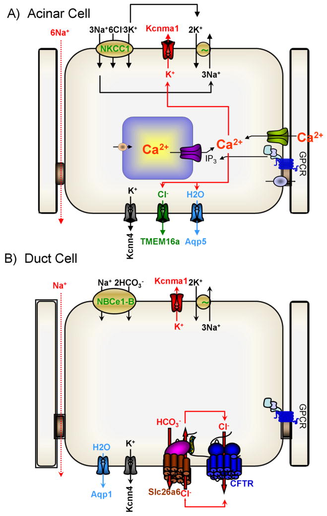
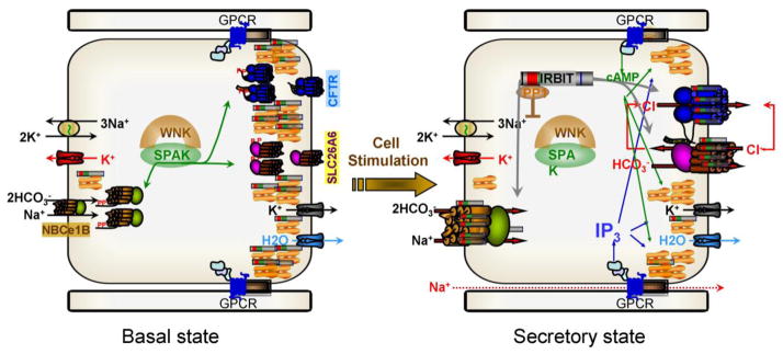
A central function of epithelia is the control of the volume and electrolyte composition of bodily fluids through vectorial transport of electrolytes and the obligatory H2O. In exocrine glands, fluid and electrolyte secretion is carried out by both acinar and duct cells, with the portion of fluid secreted by each cell type varying among glands. All acinar cells secrete isotonic, plasma-like fluid, while the duct determines the final electrolyte composition of the fluid by absorbing most of the Cl(-) and secreting HCO3 (-). The key transporters mediating acinar fluid and electrolyte secretion are the basolateral Na(+)/K(+) /2Cl(-) cotransporter, the luminal Ca(2+)-activated Cl(-) channel ANO1 and basolateral and luminal Ca(2+)-activated K(+) channels. Ductal fluid and HCO3 (-) secretion are mediated by the basolateral membrane Na(+)-HCO3 (-) cotransporter NBCe1-B and the luminal membrane Cl(-)/HCO3 (-) exchanger slc26a6 and the Cl(-) channel CFTR. The function of the transporters is regulated by multiple inputs, which in the duct include major regulation by the WNK/SPAK pathway that inhibit secretion and the IRBIT/PP1 pathway that antagonize the effects of the WNK/SPAK pathway to both stimulate and coordinate the secretion. The function of these regulatory pathways in secretory glands acinar cells is yet to be examined. An important concept in biology is synergism among signaling pathways to generate the final physiological response that ensures regulation with high fidelity and guards against cell toxicity. While synergism is observed in all epithelial functions, the molecular mechanism mediating the synergism is not known. Recent work reveals a central role for IRBIT as a third messenger that integrates and synergizes the function of the Ca(2+) and cAMP signaling pathways in activation of epithelial fluid and electrolyte secretion. These concepts are discussed in this review using secretion by the pancreatic and salivary gland ducts as model systems.


Figures

Fig. 1
Fig. 1. Mechanism of fluid and electrolyte secretion by secretory glands acinar and duct cells
The panels show the key transporters and the relationships between them that mediate the bulk of fluid and electrolyte secretion by secretory glands acinar (A) and duct (B) cells.
Fig. 2
Fig. 2. The major domains of the SLC26 transporters
The gray image is the global structure of an SLC26 transporters obtained by small angle neutron scattering (22). The core transmembrane sector is modeled based on similarity to the ClC transmembrane sector (97), and the STAS domain is the solved crystal structure of Slc26a5 (106). The images were taken from the respective references.
Fig. 3
Fig. 3. The autoinhibitory domain (AID) of NBCe1-B and a potential AIDs in slc26a6 and CFTR
Panel (a) show alignment of a sequence within NBCe1-B AID to which IRBIT and PI(4,5)P2 bind that require the conserved arginines (highlighted in red) and the homologous sequence in NBCn1-A and NDCBE-A. Panel (b) shows traces of NBCe1-B current in HeLa cells transfected with vector (blue), NBCe1-B (black) and NBCe1-B+IRBIT (red). Panel (c) shows the mea±s.e.m of the NBCe1-B current measured with the indicated mutants in the NBCe1-B AID. Panel (d) is an alignment of the NBCe1-B AID with sequences within the CFTR R domain and slc26a6 STAS domain. Panel (e) shows that IRBIT does not activate ΔR-CFTR. Panel (f) shows a model of the first 400 residues of NBCe1-B. The green domain encompasses residues 100–400 of NBCe1-B and is similar to the structure of the N terminus of AE1. Residues 40–62 are in orange and the rest in turquoise. The mutated arginines are shown in red in sticks form and the residues phosphorylated by SPAK are in blue. The results were taken from (51).
Fig. 4
Fig. 4. IRBIT mediates the synergism between PKA and Ca2+ signaling pathways
Panel (a) depict the synergistic activation of slc26a6 by 0.1μM forskolin and 0.1 and 0.3μM ATP. HeLa cells transfected with slc26a6 or slc26a6 and IRBIT(ΔPEST) were stimulated with ATP alone (close black circles), forskolin alone (open green circles) or with 0.1μM forskolin and the various concentrations of ATP (close red circles and open purple triangles). The results are the mea±s.e.m of 4–6 experiments. Panel (b) shows the synergistic activation of CFTR by 0.5μM forskolin and 3μM ATP. Maximal CFTR current is evoked by stimulation with 5 μM forskolin. Panels (c, d) show that IRBIT is required for synergistic activation of ductal fluid secretion. Fluid secretion in pancreatic ducts from wild-type and IRBIT−/− mice was measured in sealed ducts in HCO3-buffered media and stimulated with 5μM forskolin or 30nM secretin (black circles), low concentration of 0.1μM forskolin or 2nM secretin (green circles), 1μM carbachol (blue circles) and the combination of 2nM secretin and 1μM carbachol (red circles).
Fig. 5
Fig. 5. IRBIT-mediated synergistic activation of epithelial fluid and HCO3 secretion by the cAMP and Ca2+ signaling pathways
In the resting state the WNK/SPAK kinases associate with the transporters and SPAK phosphorylates NBCe1-B AID, Slc26a6 STAS domain and CFTR R domain to sequester most of them in intracellular organelles. Al low cytoplasmic IP3 IRBIT is bound to the IP3 receptors that in secretory glands are clustered at the apical pole. When the cells are stimulated with a combination of physiological concentrations of IP3 and cAMP generating agonists, PKA phosphorylates the IP3Rs to facilitate release of IRBIT from the IP3Rs by IP3 binding. IRBIT recruits PP1 to the transporters to dephosphorylate them and at the SPAK phosphorylation sites and target them to the plasma membrane. IRBIT remains bound to the transporters AIDs to relieve their constitutive inhibition resulting in activation of the transporters and of fluid and electrolyte secretion.

References

    1. Abuladze N, Lee I, Newman D, Hwang J, Boorer K, Pushkin A, Kurtz I. Molecular cloning, chromosomal localization, tissue distribution, and functional expression of the human pancreatic sodium bicarbonate cotransporter. J Biol Chem. 1998;273:17689–95. - PubMed
    1. Alexander RT, Grinstein S. Na+/H+ exchangers and the regulation of volume. Acta Physiol (Oxf) 2006;187:159–67. - PubMed
    1. Alper SL. Molecular physiology and genetics of Na+-independent SLC4 anion exchangers. J Exp Biol. 2009;212:1672–83. - PMC - PubMed
    1. Anderova M, Duchene AD, Barbara JG, Takeda K. Vasoactive intestinal peptide potentiates and directly stimulates catecholamine secretion from rat adrenal chromaffin cells. Brain Res. 1998;809:97–106. - PubMed
    1. Ando H, Mizutani A, Kiefer H, Tsuzurugi D, Michikawa T, Mikoshiba K. IRBIT suppresses IP3 receptor activity by competing with IP3 for the common binding site on the IP3 receptor. Mol Cell. 2006;22:795–806. - PubMed

Publication types