Staging TDP-43 pathology in Alzheimer's disease
- PMID: 24240737
- PMCID: PMC3944799
- DOI: 10.1007/s00401-013-1211-9
Staging TDP-43 pathology in Alzheimer's disease
Abstract
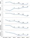
TDP-43 immunoreactivity occurs in 19-57 % of Alzheimer's disease (AD) cases. Two patterns of TDP-43 deposition in AD have been described involving hippocampus (limbic) or hippocampus and neocortex (diffuse), although focal amygdala involvement has been observed. In 195 AD cases with TDP-43, we investigated regional TDP-43 immunoreactivity with the aim of developing a TDP-43 in AD staging scheme. TDP-43 immunoreactivity was assessed in amygdala, entorhinal cortex, subiculum, hippocampal dentate gyrus, occipitotemporal, inferior temporal and frontal cortices, and basal ganglia. Clinical, neuroimaging, genetic and pathological characteristics were assessed across stages. Five stages were identified: stage I showed scant-sparse TDP-43 in the amygdala only (17 %); stage II showed moderate-frequent amygdala TDP-43 with spread into entorhinal and subiculum (25 %); stage III showed further spread into dentate gyrus and occipitotemporal cortex (31 %); stage IV showed further spread into inferior temporal cortex (20 %); and stage V showed involvement of frontal cortex and basal ganglia (7 %). Cognition and medial temporal volumes differed across all stages and progression across stages correlated with worsening cognition and medial temporal volume loss. Compared to 147 AD patients without TDP-43, only the Boston Naming Test showed abnormalities in stage I. The findings demonstrate that TDP-43 deposition in AD progresses in a stereotypic manner that can be divided into five distinct topographic stages which are supported by correlations with clinical and neuroimaging features. Given these findings, we recommend sequential regional TDP-43 screening in AD beginning with the amygdala.
Conflict of interest statement
The authors declare that they have no conflicts of interest
Figures


References
-
- Arai T, Hasegawa M, Akiyama H, et al. TDP-43 is a component of ubiquitin-positive tau-negative inclusions in frontotemporal lobar degeneration and amyotrophic lateral sclerosis. Biochem Biophys Res Commun. 2006;351:602–611. - PubMed
-
- Arai T, Mackenzie IR, Hasegawa M, et al. Phosphorylated TDP-43 in Alzheimer's disease and dementia with Lewy bodies. Acta neuropathologica. 2009;117:125–136. - PubMed
Publication types
MeSH terms
Substances
Grants and funding
LinkOut - more resources
Full Text Sources
Other Literature Sources
Medical

