Review
. 2014:800:113-31.
doi: 10.1007/978-94-007-7687-6_7.

The PAR polarity complex and cerebellar granule neuron migration

Affiliations
Review

The PAR polarity complex and cerebellar granule neuron migration

Joseph S Ramahi et al. Adv Exp Med Biol. 2014.

Abstract

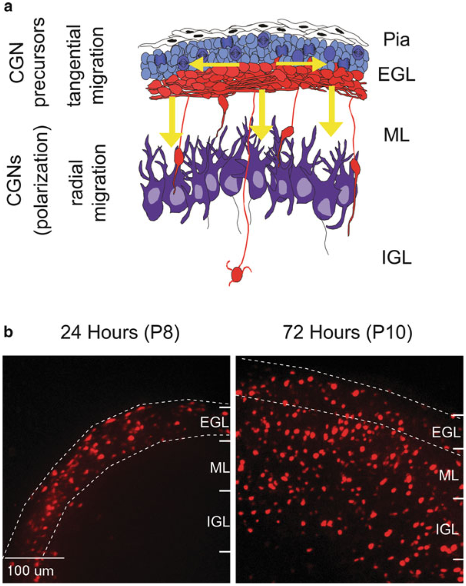
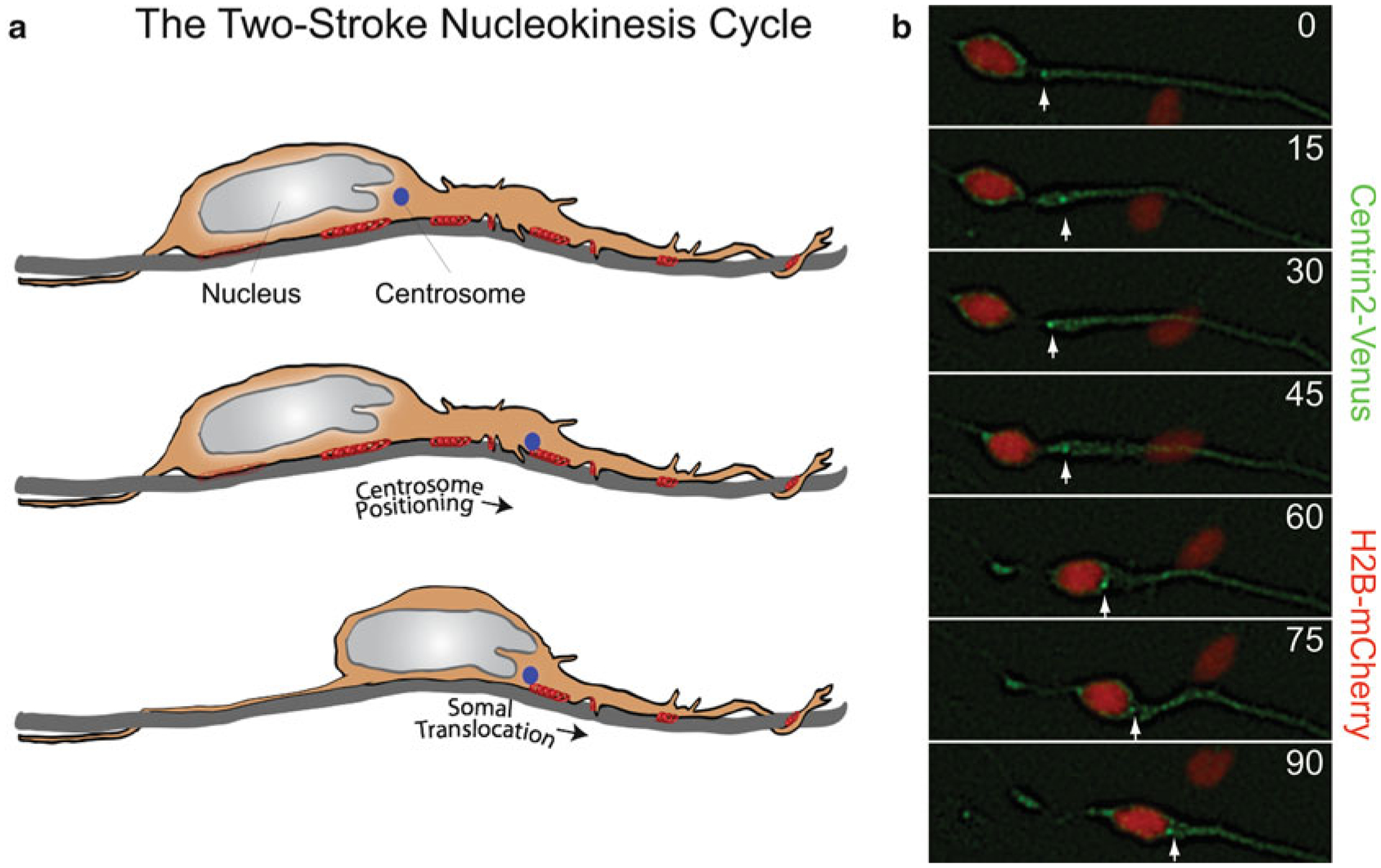
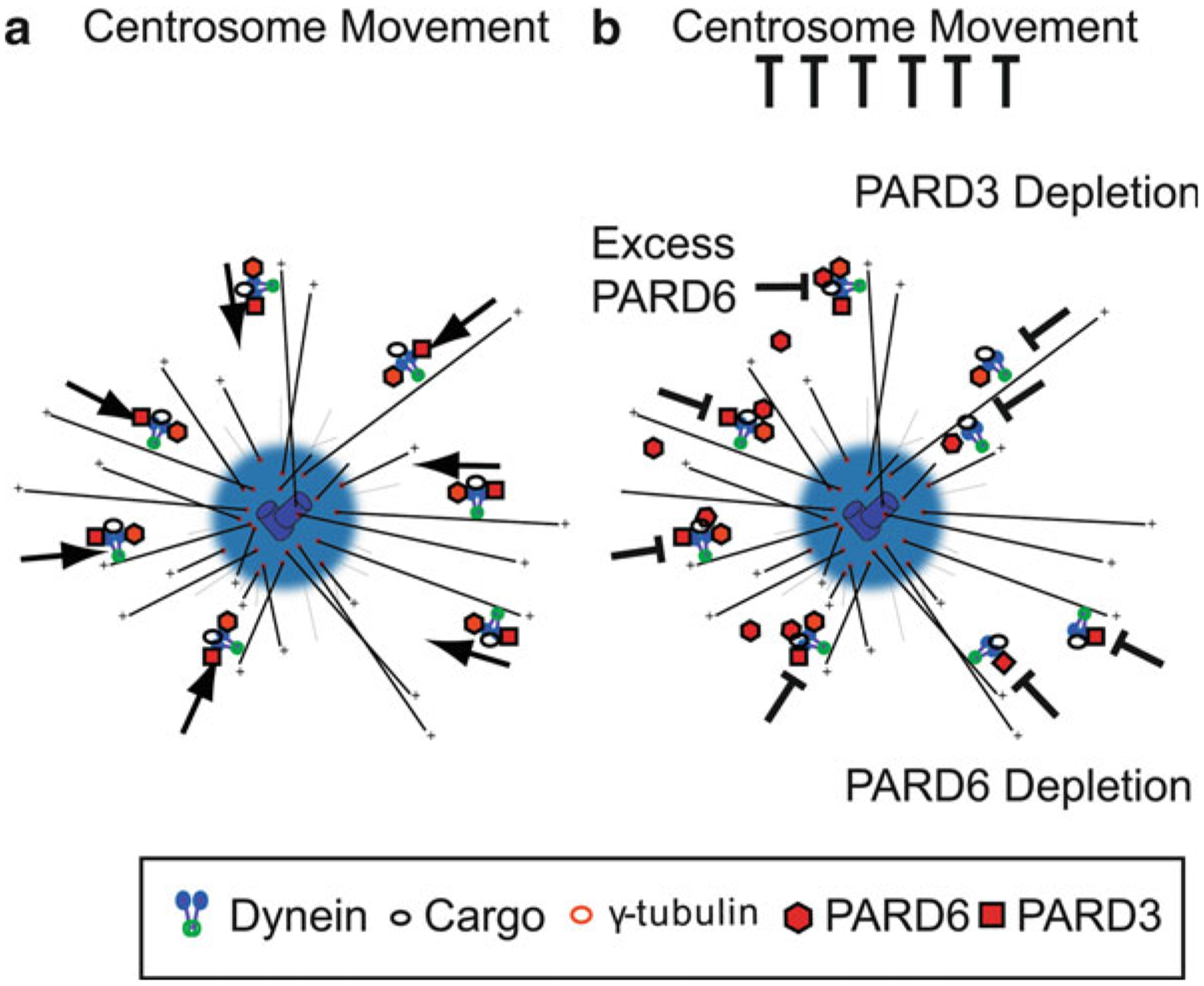
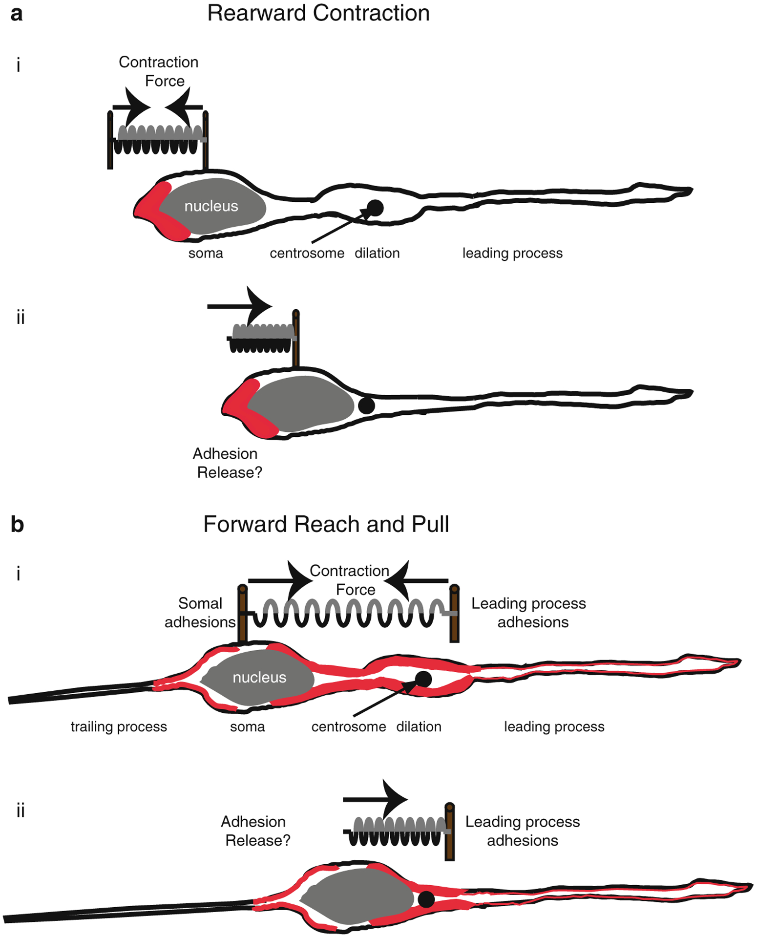
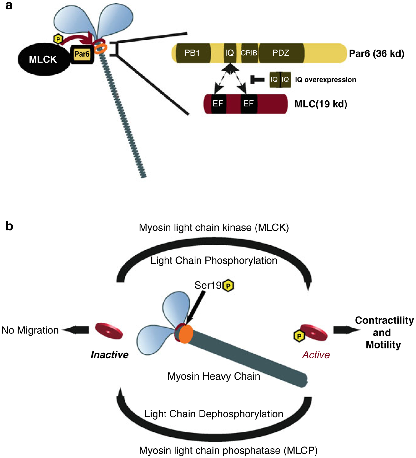
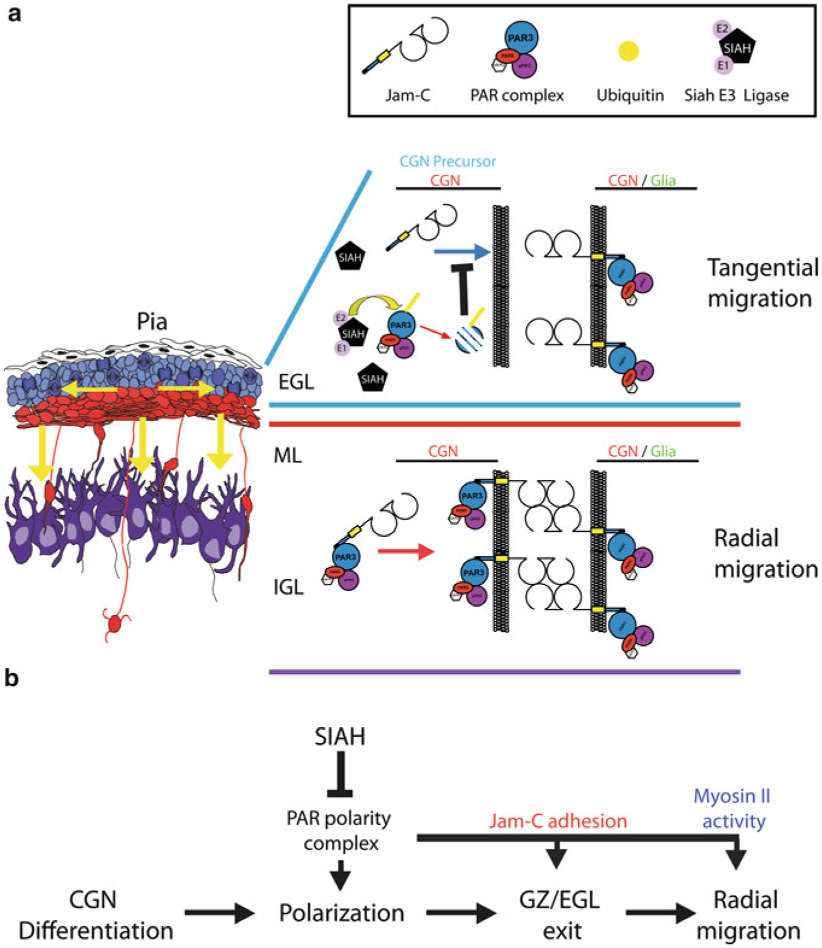
Proper migration of neurons is one of the most important aspects of early brain development. After neuronal progenitors are born in their respective germinal niches, they must migrate to their final locations to form precise neural circuits. A majority of migrating neurons move by associating and disassociating with glial fibers, which serve as scaffolding for the developing brain. Cerebellar granule neurons provide a model system for examination of the mechanisms of neuronal migration in dissociated and slice culture systems; the ability to purify these cells allows migration assays to be paired with genetic, molecular, and biochemical findings. CGNs migrate in a highly polarized fashion along radial glial fibers, using a two-stroke nucleokinesis cycle. The PAR polarity complex of PARD3, PARD6, and an atypical protein kinase C (aPKC) regulate several aspects of neuronal migration. The PAR polarity complex regulates the coordinated movements of the centrosome and soma during nucleokinesis, and also the stability of the microtubule cytoskeleton during migration. PAR proteins coordinate actomyosin dynamics in the leading process of migrating neurons, which are required for migration. The PAR complex also controls the cell-cell adhesions made by migrating neurons along glial cells, and through this mechanism regulates germinal zone exit during prenatal brain development. These findings suggest that the PAR complex coordinates the movement of multiple cellular elements as neurons migrate and that further examination of PAR complex effectors will not only provide novel insights to address fundamental challenges to the field but also expand our understanding of how the PAR complex functions at the molecular level.


Figures

Fig. 7.1
Fig. 7.1. Germinal zone migration in the developing cerebellum.
(a) Cerebellar granule neuron precursors (cGNPs) migrate tangentially (horizontal arrows) within the External Granule Layer (EGL). They then transition to a radial migration mode (vertical arrows) and migrate along glial fibers through the Molecular Layer (ML) and into the Internal Granule Layer (IGL). (b) Cereballarslice cultures electroporated with CGN-specific H2B-mCherry nuclei to track neuronal migration. At postnatal day 8 (P8, 24 h post electroporation), H2B-mCherry labeled CGNs migrate tangentially through the EGL. By P10 (72 h post electroporation) most CGNs have evacuated the EGL and migrated radially into the ML and IGL
Fig. 7.2
Fig. 7.2. The two-stroke nucleokinesis cycle of migrating neurons.
(a) In the two stroke nucleokinesis cycle, the centrosome is positioned into the neuronal leading process before somal translocation. (b) Time-lapse imaging of a migrating CGN whose centrosome is labeled with Centrin2-Venus (green) (white arrow) and whose nucleus is labeled with H2B-mCherry (red). Centrosome positioning occurs 0 and 30 min, and somal translocation occurs between 45 and 90 min
Fig. 7.3
Fig. 7.3. PAR proteins and cytoplasmic Dynein directed minus-end transport.
(a) Model of Dynein-directed minus-end transport of centrosome components mediated by PAR. This mechanism is responsible for proper centrosome motility. (b) Disruption of PAR protein components may result in inhibition of dynein mediated centrosome assembly and centrosome motility by PARD6 overexpression (Solecki et al. 2004), PARD6 RNAi (Kodani et al. 2010), and PARD3 depletion (Schmoranzer et al. 2009)
Fig. 7.4
Fig. 7.4. Actomyosin pulling models for Glial-guided neuronal migration.
(a) Rearward Contraction model. (i) Prior to somal movement, actomyosin (red) is heavily enriched at the cell rear. (ii) During somal movement myosin II squeezing at the rear is thought to “push” the cell body forward. (b) Reach and Pull model. (i) Prior to somal movement, actomyosin (red) is heavily enriched in the leading process from the cytoplasmic dilation to the neuronal soma. Given a muscle-like contraction of the F-actin array by myosin II, a taut spring effectively describes the forces produced when leading process and somal actomyosin anchoring (i.e., adhesions) are balanced before somal movement: one force vector points from the leading process back towards the soma whereas another force vector points from the soma towards the dilation (the future direction of somal movement). (ii) Once somal adhesions release, as described in (Gregory et al. 1988), actomyosin tension generated in the leading process primes somal movement towards the cytoplasmic dilation (Reproduced with permission of (Trivedi and Solecki 2011))
Fig. 7.5
Fig. 7.5. Model of Par6α interaction with the Myosin II motor complex and the Myosin cycle.
(a) Par6α binds to both MLC and MLCK, key signaling nodes regulating actomyosin contractility. Inset : The PARD6-MLC interaction may be mediated by the IQ domain of Par6α (IQ Motif (aa 104–120) = AFASNSLQRRKKGLLLRPV) and the EF hand domains of MLC. (b) Myosin contractility is dependent on Myosin Light Chain (MLC) phosphorylation by Myosin Light Chain Kinase (MLCK) at Ser19 and is required for neuronal migration. De-phosphorylation of MLC by Myosin Light Chain Phosphatase (MLCP) results in MLC inactivity and lack of myosin contractility. MCL and MLCP activity cycles Myosin contractility in migratory cells ((a) Reproduced with permission of (Solecki et al. 2009))
Fig. 7.6
Fig. 7.6. Model of SIAH E3 Ligase regulation of germinal zone exit.
(a) During cerebellar development CGN precursors migrate tangentially within the EGL. Upon differentiation and polarization, CGNs exit the GZ/EGL and migrate radially to traverse the ML and assume their final position in the IGL. Within the developing postnatal cerebellum SIAH (E3 ubiquitin ligase) is highly expressed in the EGL, where it ubiquitinates PARD3A to target it for proteasome-mediated degradation. PARD3A degradation results in inactivation of the PAR polarity complex, thereby inhibiting recruitment of the JAM-C adhesion molecule to contacts between CGNs or CGN precursors and glial cells. The absence of JAM-C-mediated adhesion prevents GZ exit by restricting the radial migration of CGN precursors. (b) The PAR polarity complex is required for differentiated CGNs to polarize, exit the GZ via JAM-C-mediated adhesion, and migrate radially via activation of the myosin II motor. SIAH negatively regulates CGN polarization, GZ exit, and radial migration by inactivating the PAR polarity complex (Reproduced with permission of (Famulski et al. 2010))

Similar articles

Cited by

References

    1. Allen KM, Gleeson JG, Shoup SM, Walsh CA (1998) A YAC contig in Xq22. 3-q23, from DXS287 to DXS8088, spanning the brain-specific genes doublecortin (DCX) and PAK3. Genomics 52(2):214–218 - PubMed
    1. Barnes AP, Polleux F (2009) Establishment of axondendrite polarity in developing neurons. Annu Rev Neurosci 32:347. - PMC - PubMed
    1. Bellion A, Baudoin JP, Alvarez C, Bornens M, Metin C (2005) Nucleokinesis in tangentially migrating neurons comprises two alternating phases: forward migration of the Golgi/centrosome associated with centrosome splitting and myosin contraction at the rear. J Neurosci 25(24):5691–5699. doi:25/24/5691 [pii] 10.1523/JNEUROSCI.1030-05.2005 - DOI - PMC - PubMed
    1. Bertet C, Sulak L, Lecuit T (2004) Myosin-dependent junction remodelling controls planar cell intercalation and axis elongation. Nature 429(6992):667–671. doi: http://www.nature.com/nature/journal/v429/n6992/suppinfo/nature02590_S1.... - PubMed
    1. Chen S, Chen J, Shi H, Wei M, Castaneda-Castellanos David R, Bultje Ronald S, Pei X, Kriegstein Arnold R, Zhang M, Shi S-H (2013) Regulation of microtubule stability and organization by mammalian Par3 in specifying neuronal polarity. Dev Cell 24(1):26–40. doi: 10.1016/j.devcel.2012.11.014 - DOI - PMC - PubMed

Publication types

MeSH terms

LinkOut - more resources