. 2013 Dec 15;191(12):6231-40.
doi: 10.4049/jimmunol.1301696. Epub 2013 Nov 15.

Optineurin insufficiency impairs IRF3 but not NF-κB activation in immune cells

Affiliations

Optineurin insufficiency impairs IRF3 but not NF-κB activation in immune cells

Ivana Munitic et al. J Immunol. .

Abstract

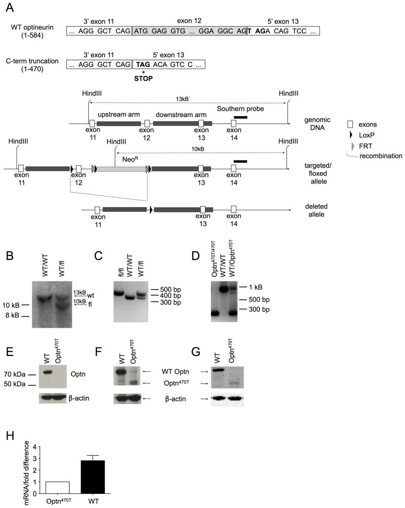
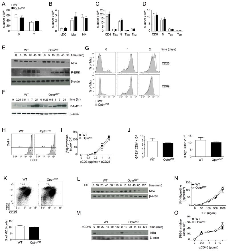
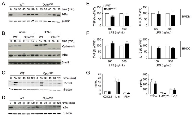
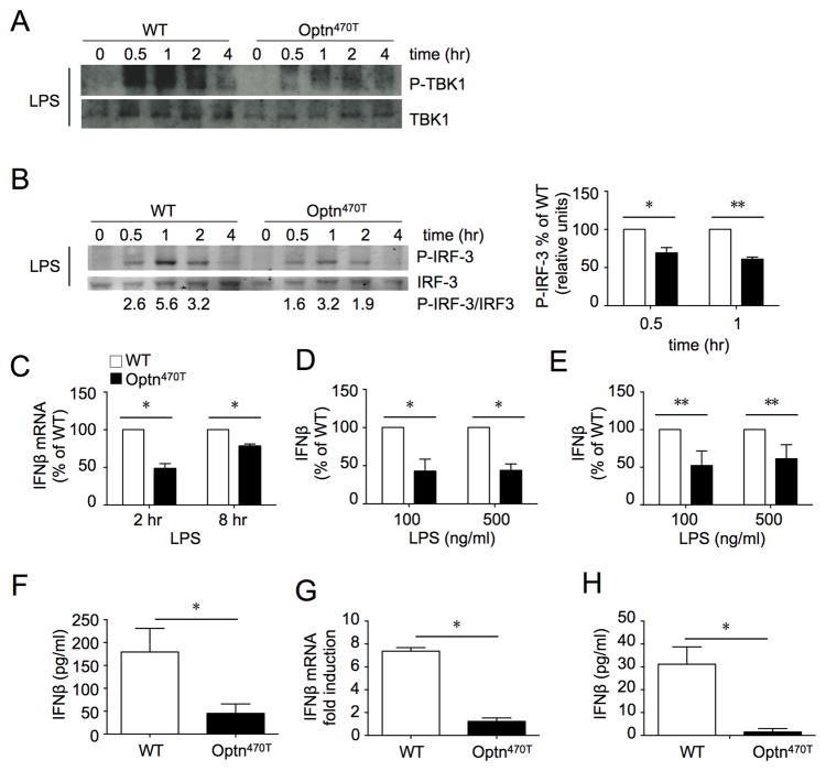
Optineurin is a widely expressed polyubiquitin-binding protein that has been implicated in regulating cell signaling via its NF-κB essential modulator-homologous C-terminal ubiquitin (Ub)-binding region. Its functions are controversial, with in vitro studies finding that optineurin suppressed TNF-mediated NF-κB activation and virus-induced activation of IFN regulatory factor 3 (IRF3), whereas bone marrow-derived macrophages (BMDMs) from mice carrying an optineurin Ub-binding point mutation had normal TLR-mediated NF-κB activation and diminished IRF3 activation. We have generated a mouse model in which the entire Ub-binding C-terminal region is deleted (Optn(470T)). Akin to C-terminal optineurin mutations found in patients with certain neurodegenerative diseases, Optn(470T) was expressed at substantially lower levels than the native protein, allowing assessment not only of the lack of Ub binding, but also of protein insufficiency. Embryonic lethality with incomplete penetrance was observed for 129 × C57BL/6 Optn(470T/470T) mice, but after further backcrossing to C57BL/6, offspring viability was restored. Moreover, the mice that survived were indistinguishable from wild type littermates and had normal immune cell distributions. Activation of NF-κB in Optn(470T) BMDM and BM-derived dendritic cells with TNF or via TLR4, T cells via the TCR, and B cells with LPS or anti-CD40 was normal. In contrast, optineurin and/or its Ub-binding function was necessary for optimal TANK binding kinase 1 and IRF3 activation, and both Optn(470T) BMDMs and bone marrow-derived dendritic cells had diminished IFN-β production upon LPS stimulation. Importantly, Optn(470T) mice produced less IFN-β upon LPS challenge. Therefore, endogenous optineurin is dispensable for NF-κB activation but necessary for optimal IRF3 activation in immune cells.


Figures

Figure 1
Figure 1. WT but not Optn470T expression inhibits activation of NF-κB and IRF3
Human optineurin constructs, homologous to murine WT (1–584), the Ub-binding point-mutant D477N, and the C-terminal truncation (1–470) are depicted (A). Human embryonic kidney 293 cells were transiently transfected with the indicated constructs and stimulated or not with TNF. Reporter luciferase activity was normalized to β-galactosidase activity (B, left panel), and anti-Flag blotting for Flag-optineurin variants is shown (B, right panel). hTLR3-293 cells, which stably express TLR3, were transiently transfected as indicated and stimulated or not with poly I:C. Luciferase activity (C, left panel), and anti-Flag blotting (C, right panel) were performed as in panel B. An average of duplicate samples of one representative experiment out of three is shown.
Figure 2
Figure 2. Generation of Optn470T mice
(A) To generate mice with C-terminal truncation of optineurin (Optn470T), a targeting construct with LoxP sites flanking exon 12 was inserted into the endogenous locus by homologous recombination. Upon Cre-mediated deletion, a stop codon was exposed at the beginning of exon 13, resulting in a truncated protein of 470 amino acids. Neo cassette, Southern probe, and recombination sites are indicated. (B) Southern blot distinguishing WT and chimeric mice (WT/fl) is shown. The introduced HindIII site in the Neo cassette leads to generation 10 kB band in the fl allele. The PCR of the indicated mice distinguishing WT and floxed (C) and WT and deleted alleles is shown (D). Blotting BMDM from the indicated mice with optineurin antibodies raised against C-terminal (E), central (F) and N-terminal epitopes (G). Optineurin mRNA was detected in BMDM by qRT-PCR, ΔCT of Optn470T was designated as 1, and the difference between ΔCT of WT and Optn470T is depicted as mean ± SEM for 3 independent experiments (H).
Figure 3
Figure 3. T and B cell development and activation are unimpaired in Optn470T mice
T and B (A), conventional DC (cDC), macrophage, and NK cell numbers (B) in spleens of the indicated mice are shown. CD4 (C) and CD8 T cell (D) numbers and their respective compartments are shown. Naïve cells were gated as CD44loCD62Lhi, TCM as CD44hiCD62Lhi, and TEM as CD44hiCD62Llo. The data from 8 mice is shown with mean ± SEM. T cells freshly purified or stimulated with anti-CD3 (3 μg/ml) and anti-CD28 (2 μg/ml) were lysed at the indicated times, and IκBα degradation and ERK1/2 phosphorylation (E), and Akt phosphorylation (F) were detected by immunoblotting. Expression of the indicated activation markers was monitored on WT and Optn470T purified T cells at the indicated times after stimulation with plate-bound anti-CD3 (3 μg/ml) and anti-CD28 (2 μg/ml) (G). CFSE-labeled T cells were stimulated as in C, and CFSE dilution was monitored after 60 hr (H). Purified T cells were cultured with 2 μg/ml of anti-CD28 and the indicated concentrations of anti-CD3 for 48 hr, pulsed with [3H]-thymidine, and harvested 18 hr later (I). WT or Optn470T mice were infected with LCMV Armstrong and the absolute numbers of GP33-tetramer+ CD8 T cells analyzed on the peak of the response (day 8; J, left panel). Splenocytes were restimulated in vitro with GP33 peptide and evaluated for cytokine production by intracellular staining. Absolute numbers of IFN-γ producing CD8 T cells are shown (J, right panel). The data represent mean ± SEM of 6 individual mice per strain from two independent experiments. Distribution of CD21hiCD23int MZ B cells (gated in B220+TCRβ- population) in splenocytes of WT and Optn470T mice was determined by flow cytometry. Numbers represent the percentage of cells in the gated region and the bar graph below represents the average of 3 mice per group ± SEM (K). B cells freshly purified or stimulated with LPS (100 ng/ml) (L) or anti-CD40 (1 μg/ml) (M) were lysed at the indicated times and IκBα was detected by immunoblotting. Purified B cells were cultured with the indicated concentration of LPS (N) or anti-CD40 (O) for 48 hr, pulsed with [3H]-thymidine, and harvested 18 hr later. Each panel is representative of two or three independent experiments.
Figure 4
Figure 4. BMDM and BMDC from Optn470T mice have unimpaired NF-κB responses
BMDM from WT and Optn470T mice were stimulated with TNF for the indicated times and degradation of IκBα was detected by immunoblotting (A). BMDM pretreated overnight with 300 U/ml of IFN-β or not were stimulated with TNF as indicated, and lysates were blotted for optineurin and IκBα (B). Cell lysates of TNF-treated BMDM were blotted for P-ERK (C). Cell lysates of LPS-stimulated BMDM were blotted for IκBα (D). A representative example for two to three experiments is shown for all blots, with β-actin staining as a loading control. The indicated cytokines secreted upon LPS stimulation of Optn470T BMDM (E) and BMDC (F) are depicted as the percent of WT values. The data from for two to three independent experiments are shown as mean ± SEM. Cytokines detected in the sera of LPS-injected WT and Optn470T BMDM are shown (G). The mean ± SEM from 4 mice from two independent experiments is shown.
Figure 5
Figure 5. BMDM and BMDC from Optn470T mice have diminished IRF3 responses
Phospho-TBK1, total TBK1 (A), and phospho-IRF3 and total IRF3 (B) were detected by blotting lysates of BMDM from WT and Optn470T mice at the indicated times after LPS treatment. The lanes were rearranged for clarity. A representative blot of 5 experiments is shown. The densitometry was done on two phospho-IRF3 and total IRF3 blots that were developed with fluorescently-labeled secondary antibodies; numbers below the blot represent the quantitation, and an average of two independent experiments ± SD is displayed (right panel). mRNA for IFN-β was amplified from BMDM at 2 and 8 hr (C), and IFN-β was measured in supernatants at 24 hr (D) after LPS treatment. The mean ± SEM of Optn470T is depicted as percent of WT. IFN-β in supernatants of WT and Optn470T BMDC treated with LPS (E). The data are representative of 2–5 experiments each. IFN-β was measured in the sera of WT and Optn470T mice 6 hr after LPS injection (F). An average of seven mice ± SEM is shown per each genotype. BMDM from WT and Optn470T mice were stimulated with CpGA (2 μM). mRNA for IFN-β was amplified after 8 hr (G), and IFN-β was measured in supernatants at 24 hr (H). The data represent mean ± SD measured from two independent experiments. * p < 0.05; ** p <0.001.

References

    1. Ghosh S, Hayden MS. Celebrating 25 years of NF-kappaB research. Immunol Rev. 2012;246:5–13. - PMC - PubMed
    1. Lebrun-Julien F, Duplan L, Pernet V, Osswald I, Sapieha P, Bourgeois P, Dickson K, Bowie D, Barker PA, Di Polo A. Excitotoxic death of retinal neurons in vivo occurs via a non-cell-autonomous mechanism. J Neurosci. 2009;29:5536–5545. - PMC - PubMed
    1. Sen R, Smale ST. Selectivity of the NF-{kappa}B response. Cold Spring Harb Perspect Biol. 2010;2:a000257. - PMC - PubMed
    1. Gerondakis S, Siebenlist U. Roles of the NF-kappaB pathway in lymphocyte development and function. Cold Spring Harb Perspect Biol. 2010;2:a000182. - PMC - PubMed
    1. Fitzgerald KA, McWhirter SM, Faia KL, Rowe DC, Latz E, Golenbock DT, Coyle AJ, Liao SM, Maniatis T. IKKepsilon and TBK1 are essential components of the IRF3 signaling pathway. Nat Immunol. 2003;4:491–496. - PubMed

Publication types

MeSH terms