

Nodular lymphocyte predominant hodgkin lymphoma and T cell/histiocyte rich large B cell lymphoma--endpoints of a spectrum of one disease?
- PMID: 24244368
- PMCID: PMC3823948
- DOI: 10.1371/journal.pone.0078812
Nodular lymphocyte predominant hodgkin lymphoma and T cell/histiocyte rich large B cell lymphoma--endpoints of a spectrum of one disease?
Abstract
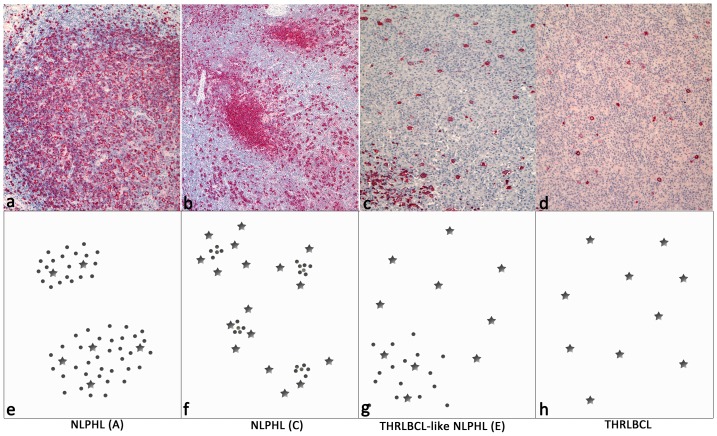
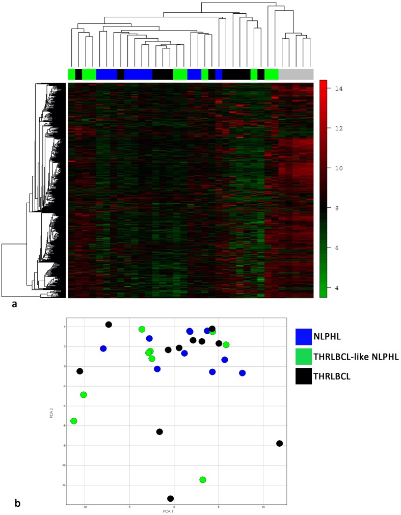
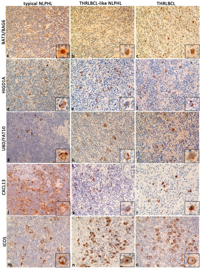
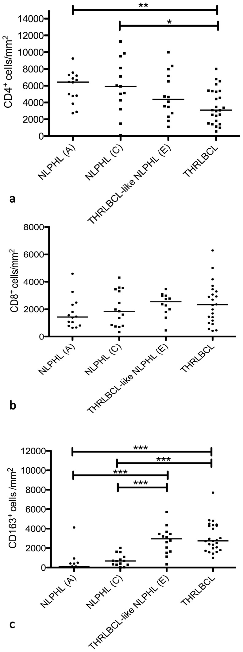
In contrast to the commonly indolent clinical behavior of nodular lymphocyte predominant Hodgkin lymphoma (NLPHL), T cell/histiocyte rich large B cell lymphoma (THRLBCL) is frequently diagnosed in advanced clinical stages and has a poor prognosis. Besides the different clinical presentations of these lymphoma entities, there are variants of NLPHL with considerable histopathologic overlap compared to THRLBCL. Especially THRLBCL-like NLPHL, a diffuse form of NLPHL, often presents a histopathologic pattern similar to THRLBCL, suggesting a close relationship between both lymphoma entities. To corroborate this hypothesis, we performed gene expression profiling of microdissected tumor cells of NLPHL, THRLBCL-like NLPHL and THRLBCL. In unsupervised analyses, the lymphomas did not cluster according to their entity. Moreover, even in supervised analyses, very few consistently differentially expressed transcripts were found, and for these genes the extent of differential expression was only moderate. Hence, there are no clear and consistent differences in the gene expression of the tumor cells of NLPHL, THRLBCL-like NLPHL and THRLBCL. Based on the gene expression studies, we identified BAT3/BAG6, HIGD1A, and FAT10/UBD as immunohistochemical markers expressed in the tumor cells of all three lymphomas. Characterization of the tumor microenvironment for infiltrating T cells and histiocytes revealed significant differences in the cellular composition between typical NLPHL and THRLBCL cases. However, THRLBCL-like NLPHL presented a histopathologic pattern more related to THRLBCL than NLPHL. In conclusion, NLPHL and THRLBCL may represent a spectrum of the same disease. The different clinical behavior of these lymphomas may be strongly influenced by differences in the lymphoma microenvironment, possibly related to the immune status of the patient at the timepoint of diagnosis.
Conflict of interest statement
Figures
References
-
- Küppers R, Rajewsky K, Zhao M, Simons G, Laumann R, et al. (1994) Hodgkin disease: Hodgkin and Reed-Sternberg cells picked from histological sections show clonal immunoglobulin gene rearrangements and appear to be derived from B cells at various stages of development. Proc Natl Acad Sci USA 91: 10962–10966. - PMC - PubMed
-
- Diehl V, Sextro M, Franklin J, Hansmann ML, Harris N, et al. (1999) Clinical presentation, course, and prognostic factors in lymphocyte-predominant Hodgkin’s disease and lymphocyte-rich classical Hodgkin’s disease: report from the European Task Force on Lymphoma Project on Lymphocyte-Predominant Hodgkin’s Disease. J Clin Oncol 17: 776–783. - PubMed
-
- Swerdlow SH, International Agency for Research on Cancer, World Health Organization (2008) WHO classification of tumours of haematopoietic and lymphoid tissues. Lyon, France: International Agency for Research on Cancer. 439 p. p.
-
- Fan Z, Natkunam Y, Bair E, Tibshirani R, Warnke RA (2003) Characterization of variant patterns of nodular lymphocyte predominant hodgkin lymphoma with immunohistologic and clinical correlation. Am J Surg Pathol 27: 1346–1356. - PubMed
Publication types
MeSH terms
Substances
LinkOut - more resources
Full Text Sources
Other Literature Sources
Medical
Molecular Biology Databases
