. 2013 Nov 14;8(11):e79255.
doi: 10.1371/journal.pone.0079255. eCollection 2013.

KSRP modulation of GAP-43 mRNA stability restricts axonal outgrowth in embryonic hippocampal neurons

Affiliations

KSRP modulation of GAP-43 mRNA stability restricts axonal outgrowth in embryonic hippocampal neurons

Clark W Bird et al. PLoS One. .

Abstract

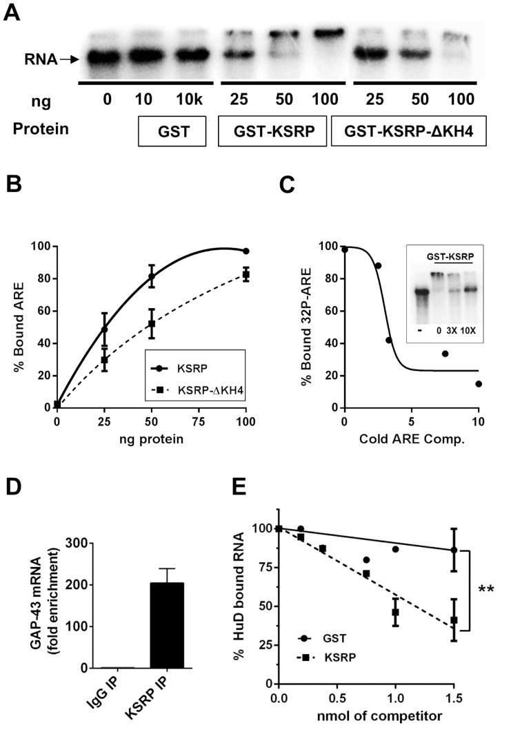
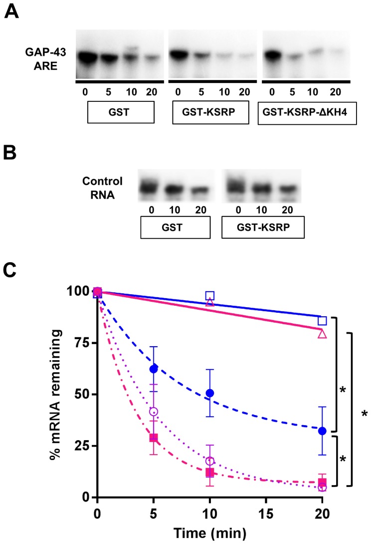
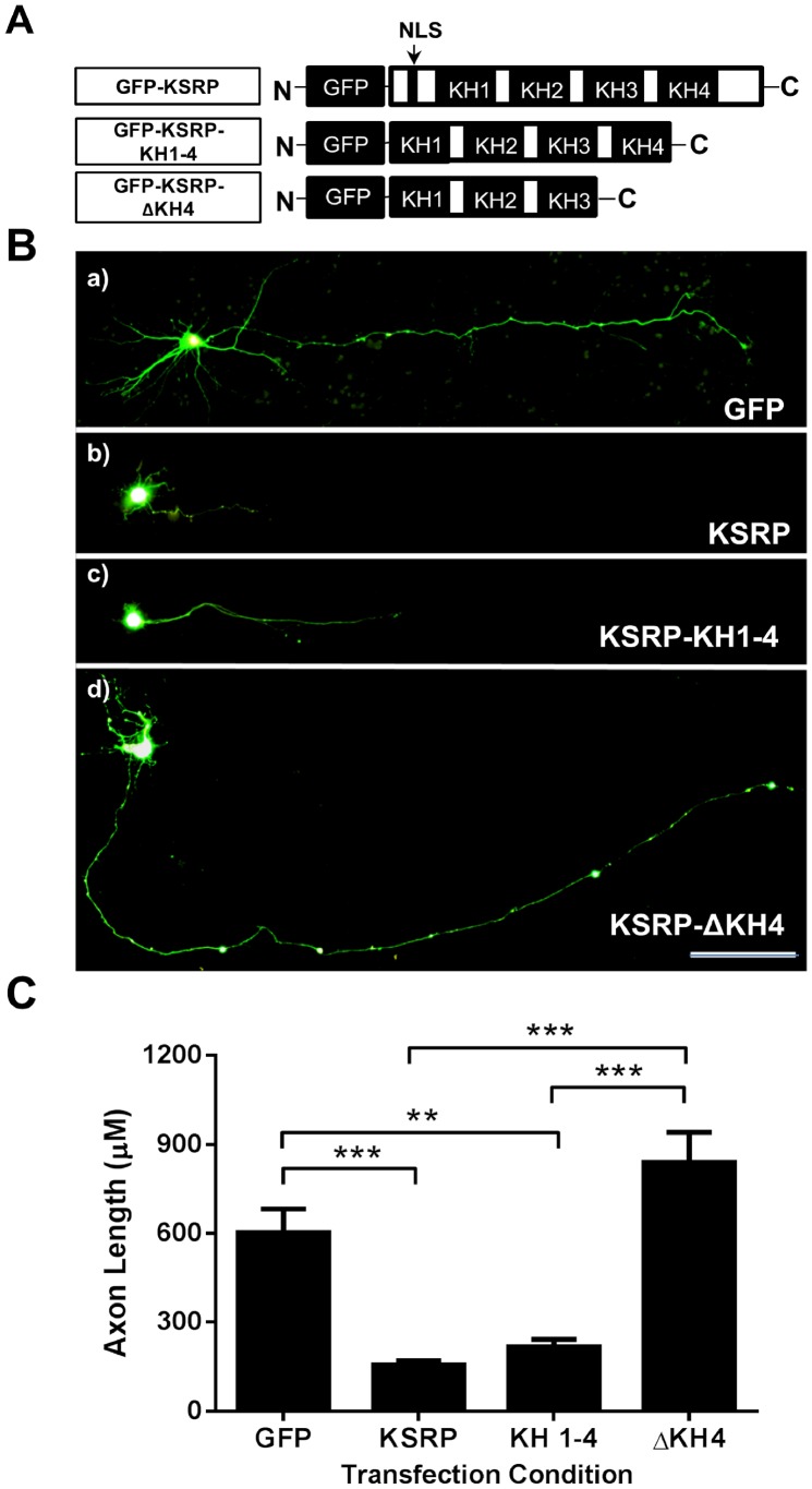
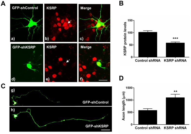
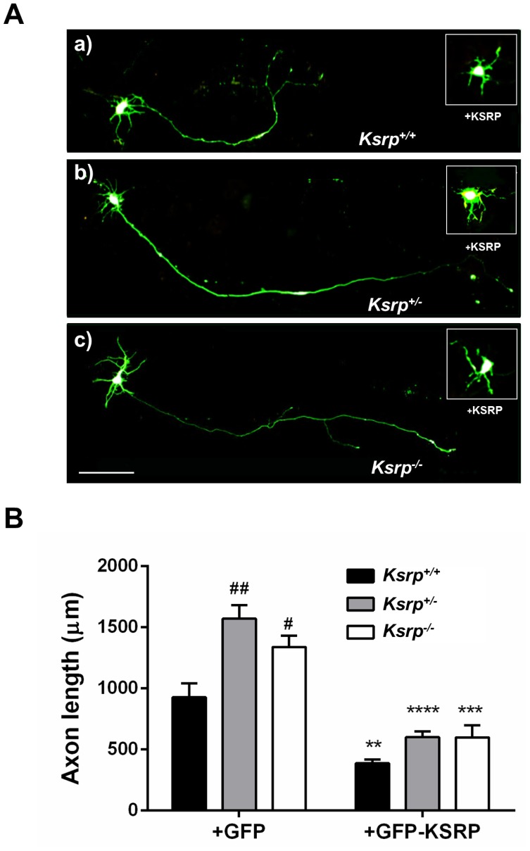
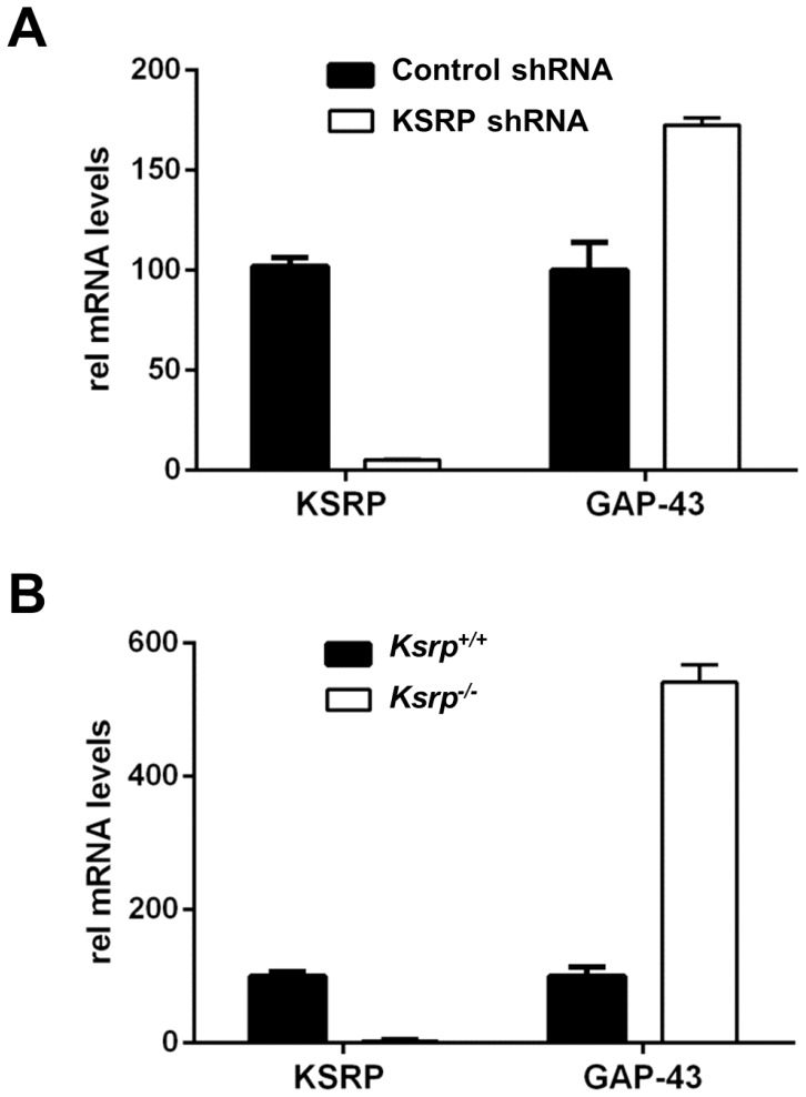
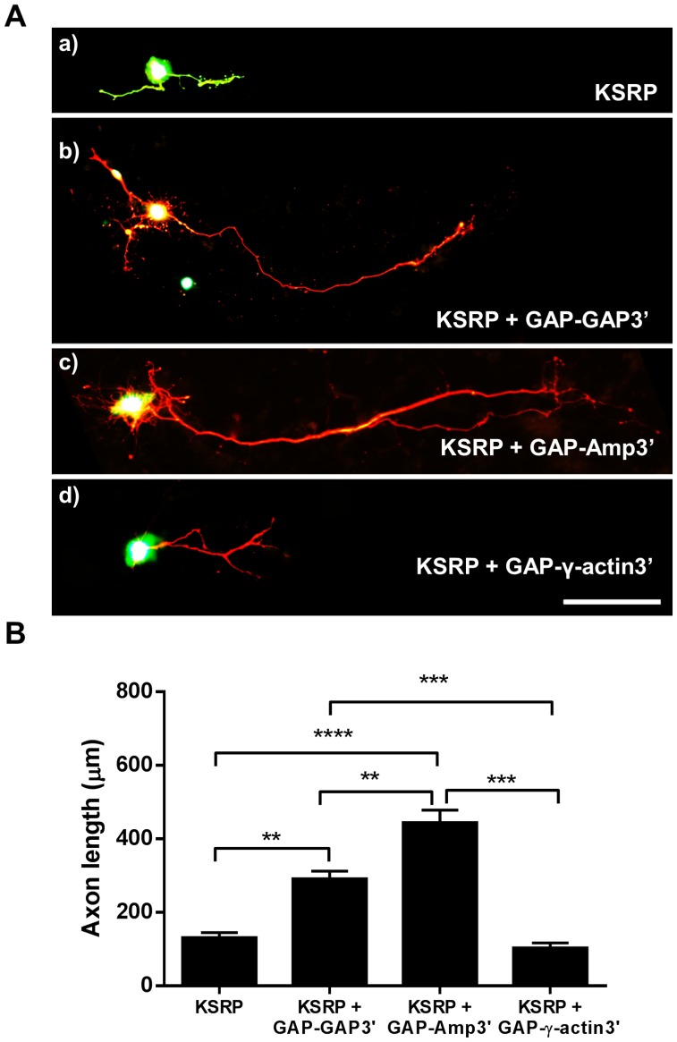
The KH-type splicing regulatory protein (KSRP) promotes the decay of AU-rich element (ARE)-containing mRNAs. Although KSRP is expressed in the nervous system, very little is known about its role in neurons. In this study, we examined whether KSRP regulates the stability of the ARE-containing GAP-43 mRNA. We found that KSRP destabilizes this mRNA by binding to its ARE, a process that requires the presence of its fourth KH domain (KH4). Furthermore, KSRP competed with the stabilizing factor HuD for binding to these sequences. We also examined the functional consequences of KSRP overexpression and knockdown on the differentiation of primary hippocampal neurons in culture. Overexpression of full length KSRP or KSRP without its nuclear localization signal hindered axonal outgrowth in these cultures, while overexpression of a mutant protein without the KH4 domain that has less affinity for binding to GAP-43's ARE had no effect. In contrast, depletion of KSRP led to a rise in GAP-43 mRNA levels and a dramatic increase in axonal length, both in KSRP shRNA transfected cells and neurons cultured from Ksrp(+/-) and Ksrp(-/-) embryos. Finally we found that overexpression of GAP-43 rescued the axonal outgrowth deficits seen with KSRP overexpression, but only when cells were transfected with GAP-43 constructs containing 3' UTR sequences targeting the transport of this mRNA to axons. Together, our results suggest that KSRP is an important regulator of mRNA stability and axonal length that works in direct opposition to HuD to regulate the levels of GAP-43 and other ARE-containing neuronal mRNAs.


Conflict of interest statement

Competing Interests: The authors want to clarify that all the funding for this work came from NIH grants or a small private foundation and none of the authors have any conflicts of interest. Specifically, the authors want to assure PLOS ONE that Dr. Bolognani worked on this manuscript while he was a post-doc in the authors' lab before joining Roche Pharmaceuticals as a staff scientist. This does not alter the authors' adherence to all the PLOS ONE policies on sharing data and materials.

Figures

Figure 1
Figure 1. Binding of KSRP to GAP-43 ARE.
A. REMSA assays with increasing amounts of purified GST, GST-KSRP, or GST-KSRP-ΔKH4 protein and 32P-labeled GAP-43 ARE. Arrow shows the migration of unbound RNA B. Binding curves comparing the relative affinities of the GST-KSRP and GST-KSRP-ΔKH4 for the GAP-43 ARE. Results are the average of 3 separate experiments. C. Displacement of bound radiolabeled GAP-43 ARE from GST-KSRP in the presence of excess of cold competitor. D. Enrichment of GAP-43 mRNA precipitated using KSRP antibodies vs. control IgG following in vivo UV-crosslinking immunoprecitation (CLIP) assays. E. Competitive binding assay of HuD and KSRP. 32P-labeled GAP-43 ARE was incubated with HuD and increasing amounts of either GST or KSRP competitor protein, before HuD was pulled down and bound RNA measured by scintillation counting. **p<0.01 for difference between the two lines (GST, y = −10x+100 vs. KSRP, y = −44x+100) by Fisher’s r-to-z-test.
Figure 2
Figure 2. KSRP increases GAP-43 ARE decay in vitro.
A. Representative images from mRNA decay assays. Purified recombinant GST, GST-KSRP, or GST-KSRP-ΔKH4 protein was incubated with 32P-labeled GAP-43 ARE in the presence of S100 extracts from Ksrp−/− mice. B. In vitro decay of a control GAP-43 RNA that does not contain the ARE and is stable even in the presence of KSRP. C. Decay curves showing the results of 3 separate decay experiments fitted with a single rate exponential decay curve. Comparisons of the different decay rates were performed by Two-Way ANOVAs after transforming the data to ln: *p<0.05 ARE/GST vs. ARE/GST-KSRP[F (1, 20) = 4.65333, p = 0.04334]; ARE/GST vs.Co/GST [F (1,14) = 6.2256, p = 0.02571] and ARE/GST-KSRP vs. Co/GST-KSRP [F (1,14) = 6.88683, p = 0.02001].
Figure 3
Figure 3. Overexpression of KSRP limits axonal outgrowth in hippocampal neurons.
A. GFP-KSRP constructs cloned for KSRP overexpression studies. GFP-KSRP-KH 1–4 lacks a NLS, while the GFP-KSRP-ΔKH4 lacks both a NLS and the fourth KH domain. B. Rat hippocampal neurons transfected with KSRP constructs. Transfection conditions: a) GFP, b) KSRP, c) KSRP KH 1–4, d) KSRP ΔKH4. Scale bar is 100 µm. C. Quantitation of axonal outgrowth in KSRP transfected hippocampal neurons. Averaged axonal outgrowth from several transfection experiments is shown (mean +/− SEM). **, p<0.01; ***p<0.001 using a One way ANOVA with Tukey’s multiple comparison post-hoc tests (GFP n = 8, KSRP n = 11, KSRP KH 1–4 n = 6, KSRP ΔKH4 n = 10).
Figure 4
Figure 4. shRNA knockdown of KSRP increases axonal length in transfected hippocampal neurons.
A. Representative images of shRNA transfections. Control (a–c) or KSRP (d–f) shRNA plasmids were transfected into primary hippocampal neuronal cultures, and KSRP expression was quantified by comparing KSRP immunofluorescence levels with untransfected cells in the same field. Arrows point to the nuclei of transfected cells. Scale bar is 25 µm. B. Quantification of KSRP knockdown by GFP-KSRP-shRNA. Knockdown was measured by comparing KSRP immunofluorescence intensity in the nuclei of shRNA transfected cells vs. untransfected cells in the same image frame. Plotted graphs are relative mean (+/− SEM) levels of KSRP fluorescence intensity. **, p<0.01, Student’s t-test (n = 7 for control shRNA; n = 14 for KSRP shRNA). C. Rat hippocampal neurons transfected with shRNA constructs. Transfection conditions: g) Non-targeting GFP-shRNA control vector or h) GFP-KSRP-shRNA. Scale bar is 100 µm. D. Quantitation of axonal outgrowth in shRNA transfected hippocampal neurons. Averaged axonal outgrowth from 10 separate transfection experiments is shown (mean +/− SEM). **p<0.01 using Student’s t-test.
Figure 5
Figure 5. Increased axonal outgrowth in Ksrp+/− and Ksrp−/− neurons is hampered by KSRP overexpression.
A. Representative images of E17 cultured hippocampal neurons from wild type (WT), KSRP heterozygous and KO embryos transfected with either control GFP (a–c) or GFP-KSRP (insets in a–c) plasmids. Scale bar is 100 µm for both main panels and insets. B. Average axonal length (mean +/− SEM) from 2–3 separate cultures of 7–9 embryos in each genotype transfected with either GFP or GFP-KSRP (n = 10–15 cells per culture). Two way ANOVA results: interaction [F (2, 26) = 2.961, p = 0.0694], genotype [F (1, 26) = 111.9, p<0.0001], and transfection condition, [F (2, 26) = 12.69, p = 0.0001]. One way ANOVA and Tukey’s multiple comparison tests demonstrate effect of genotype ## p<0.01 Ksrp+/− vs. Ksrp+/+, and # p<0.05 Ksrp−/− vs. Ksrp+/+, and effect of GFP-KSRP transfection **p<0.01, ****p<0.0001 and ***p<.0.001 for Ksrp+/+, Ksrp+/− and Ksrp−/−, respectively.
Figure 6
Figure 6. Increased levels of GAP-43 mRNA in PC12 cells with reduced KSRP expression and in E17 cortices from Ksrp−/− mice.
A. KSRP mRNA knockdown and GAP-43 upregulation in PC12 cells transfected with pGFP-shKSRP relative to control non-targeting pGFP-shRNA. GFP expressing transfected PC12 cells were enriched by FACS to collect the brightest 3% fraction of cells before using them for KSRP and GAP-43 qRT-PCR. B. RNA was extracted from E17 Ksrp+/− and Ksrp−/− cortical tissue. Ksrp−/− cortices contained significantly greater levels of GAP-43 mRNA.
Figure 7
Figure 7. Overexpression of GAP-43 rescues limited axonal outgrowth in KSRP transfected hippocampal neurons.
A. Rat hippocampal neurons transfected with GFP-KSRP and various mCherry-GAP-43 (mCherry-GAP) constructs with different 3′ UTRs. Transfection conditions: a) GFP-KSRP only, b) GFP-KSRP+mCherry-GAP-GAP3’, c) GFP-KSRP+mCherry-GAP-Amp3’, d) GFP-KSRP+mCherry-GAP-γ-actin3. Scale bar is 100 µM. B. Quantitation of axonal outgrowth in transfected neurons. Averaged data from at least 5 neurons per transfection from 6 separate transfection experiments is shown (mean +/− SEM). **p<0.01, ***p<0.001 and ****p<0.0001 using One way ANOVA with Tukey’s multiple comparison post-hoc tests.

References

    1. Eberwine J, Miyashiro K, Kacharmina JE, Job C (2001) Local translation of classes of mRNAs that are targeted to neuronal dendrites. Proc Natl Acad Sci U S A 98: 7080–7085. - PMC - PubMed
    1. Steward O, Schuman EM (2001) Protein synthesis at synaptic sites on dendrites. Annu Rev Neurosci 24: 299–325. - PubMed
    1. Martin KC, Zukin RS (2006) RNA trafficking and local protein synthesis in dendrites: an overview. J Neurosci 26: 7131–7134. - PMC - PubMed
    1. Yoo S, van Niekerk EA, Merianda TT, Twiss JL (2010) Dynamics of axonal mRNA transport and implications for peripheral nerve regeneration. Exp Neurol 223: 19–27. - PMC - PubMed
    1. Bolognani F, Perrone-Bizzozero NI (2008) RNA-protein interactions and control of mRNA stability in neurons. J Neurosci Res 86: 481–489. - PubMed

Publication types

MeSH terms