. 2013 Nov 7;8(11):e79321.
doi: 10.1371/journal.pone.0079321. eCollection 2013.

AcSDKP regulates cell proliferation through the PI3KCA/Akt signaling pathway

Affiliations

AcSDKP regulates cell proliferation through the PI3KCA/Akt signaling pathway

Ping Hu et al. PLoS One. .

Abstract

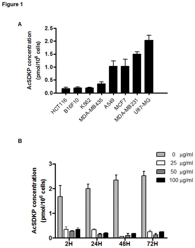
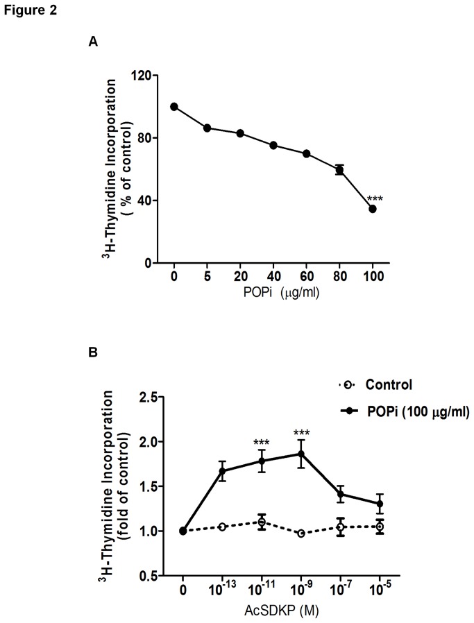
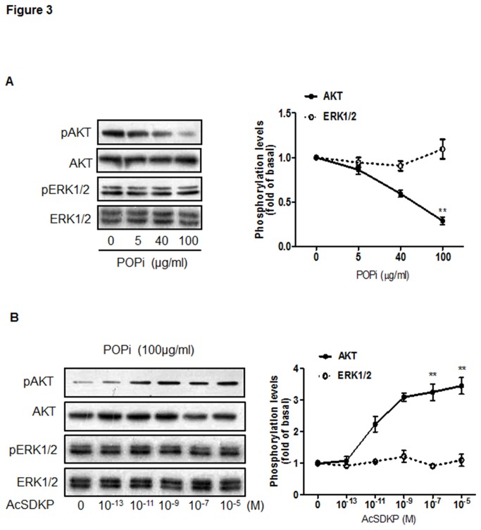
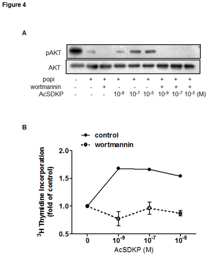
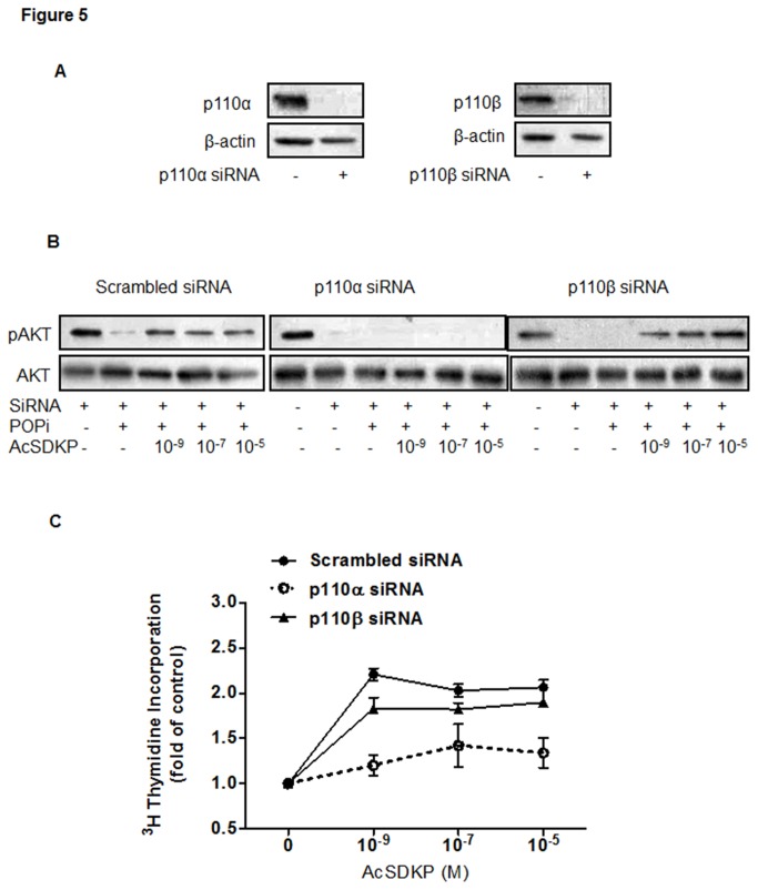
The natural tetrapeptide acetyl-N-Ser-Asp-Lys-Pro (AcSDKP) is generated from the N-terminus of thymosin-β4 through enzymatic cleavage by prolyl oligopeptidase (POP). AcSDKP regulation of proliferation of different cells is implicated in hematopoiesis and angiogenesis. This tetrapeptide present in almost all cells was recently detected at elevated concentrations in neoplastic diseases. However, previously reported in vitro and in vivo studies indicate that AcSDKP does not contribute to the pathogenesis of cancers. Here we show that exogenous AcSDKP exerts no effect on the proliferation of actively dividing malignant cells. Using S17092, a specific POP inhibitor (POPi), to suppress the biosynthesis of AcSDKP in U87-MG glioblastoma cells characterized by high intracellular levels of this peptide, we found that all tested doses of POPi resulted in an equally effective depletion of AcSDKP, which was not correlated with the dose-dependent decreases in the proliferation rate of treated cells. Interestingly, addition of exogenous AcSDKP markedly reversed the reduction in the proliferation of U87-MG cells treated with the highest dose of POPi, and this effect was associated with activation of the phosphatidylinositol-3 kinase (PI3K)/Akt pathway. However, extracellular-regulated protein kinase (ERK) activation was unaltered by S17092 and AcSDKP co-treatment. Knockdown of individual PI3K catalytic subunits revealed that p110α and p110β contributed differently to AcSDKP regulation of U87-MG cell proliferation. Disruption of p110α expression by small interfering RNA (siRNA) abrogated AcSDKP-stimulated Akt phosphorylation, whereas knockdown of p110β expression exhibited no such effect. Our findings indicate for the first time that the PI3KCA/Akt pathway mediates AcSDKP regulation of cell proliferation and suggest a role for this ubiquitous intracellular peptide in cell survival.


Conflict of interest statement

Competing Interests: The authors have declared that no competing interests exist.

Figures

Figure 1
Figure 1. Intracellular concentration of AcSDKP.
The level of endogenous AcSDKP peptide was measured daily in cells grown in 25-cm2 flasks in the absence or presence of POP inhibitor (POPi). Following trypsin detachment, the cells were counted, washed with PBS, sonicated, and AcSDKP was extracted with methanol according to the procedure described in “Materials and Methods”. (A) The AcSDKP expression in different cell lines: HCT116: human colon cancer cell; B16F10: mouse melanoma cell; K562: human leukemia cell; MCF7, MDA-MB231 and MDA-MB435: human breast cancer cell; A549: human lung cancer cell; U87-MG: human glioblastoma cell. (B) Kinetics of down-regulation by POPi of AcSDKP in U87-MG cells. Cells were treated by S17092 compound, an inhibitor of AcSDKP biosynthesis, at different concentrations from 2 to 72 h. The results represent the mean ± SEM from three experiments.
Figure 2
Figure 2. Effect of AcSDKP on proliferation of U87-MG cells in the absence or presence of S17092.
(A) Effect of S17092 on the proliferation rate of U87-MG cells. DNA synthesis was measured by [3H]-thymidine incorporation assay as described in “Materials and Methods”. Cells were treated with S17092 at different concentrations for 24 h. (B) Cells were pretreated with 100 μg/ml of S17092 for 2 h and then combined with AcSDKP at the indicated concentrations for 24 h. The data represent least-squares mean ± SEM. Values that differ significantly from untreated cells are indicated by asterisks (***p<0.001).
Figure 3
Figure 3. AcSDKP restored the proliferation of U87-MG cells through Akt activation.
(A) Effect of S17092 on the phosphorylation of Akt (Ser473) and ERK1/2 in U87-MG cells. Cells were incubated with different concentrations of S17092 for 2 h and analyzed by Western blot with antibodies against p-Akt and p-ERK1/2 proteins. (B) Effect of AcSDKP on the phosphorylation of Akt and ERK1/2 in S17092-treated U87-MG cells. Cells were pretreated with 100 μg/ml of S17092 for 2 h and then combined with AcSDKP at the indicated concentrations for 2 h. The graph represents the relative level of Akt and ERK1/2 phosphorylation compared with the control group. Similar results were obtained from three independent experiments. The ratio of band intensities was calculated after normalizing the phospho-protein signals with the total protein signals. The calculations were done using NIH image software.
Figure 4
Figure 4. Effect of wortmannin, a PI3K inhibitor, on AcSDKP-induced Akt phosphorylation and U87-MG proliferation.
Cells were pretreated with 100 μg/ml S17092 for 90 min and then with 10 μM wortmannin for additional 30 min before addition of different concentrations of AcSDKP in the presence of 100 μg/ml S17092. (A) Effect of wortmannin after co-treatment for 2 h with POPi and AcSDKP was analyzed by Western blot for p-Akt and Akt proteins. (B) Cell proliferation was determined after co-treatment for 24 h with POPi and AcSDKP by [3H]-thymidine incorporation assays performed in triplicate. Representative results are shown from three independent experiments.
Figure 5
Figure 5. AcSDKP activated Akt phosphorylation via p110α but not p110β.
(A) RNA interference effectively reduced PI3K p110α and p110β protein expression in U87-MG cells. At 4 days post transfection, activation of Akt was evaluated by Western blot. β-actin was used as the loading control. (B). Extracts from U87-MG cells transfected with p110 siRNA or scrambled RNA control were analyzed by Western blot for p-Akt and Akt proteins after co-treatment with POPi and AcSDKP as indicated. Representative results are shown from three independent experiments. (C) The effect of AcSDKP on proliferation of cells transfected with p110 siRNA or scrambled RNA control. Cell proliferation was determined by [3H]-thymidine incorporation assays performed in triplicate.

References

    1. Cavasin MA, Rhaleb NE, Yang XP, Carretero OA (2004) Prolyl oligopeptidase is involved in release of the antifibrotic peptide Ac-SDKP. Hypertension 43: 1140-1145. - PMC - PubMed
    1. Lenfant M, Wdzieczak-Bakala J, Guittet E, Prome JC, Sotty D et al. (1989) Inhibitor of hematopoietic pluripotent stem cell proliferation: purification and determination of its structure. Proc Natl Acad Sci U S A 86: 779-782. doi:10.1073/pnas.86.3.779. PubMed: 2915977. - DOI - PMC - PubMed
    1. Rhaleb NE, Peng H, Harding P, Tayeh M, LaPointe MC et al. (2001) Effect of N-acetyl-seryl-aspartyl-lysyl-proline on DNA and collagen synthesis in rat cardiac fibroblasts. Hypertension 37: 827-832. doi:10.1161/01.HYP.37.3.827. PubMed: 11244003. - DOI - PMC - PubMed
    1. Pokharel S (2002) N-Acetyl-Ser-Asp-Lys-Pro Inhibits Phosphorylation of Smad2 in Cardiac Fibroblasts. Hypertension 40: 155-161. doi:10.1161/01.HYP.0000025880.56816.FA. PubMed: 12154106. - DOI - PubMed
    1. Kanasaki K, Haneda M, Sugimoto T, Shibuya K, Isono M et al. (2006) N-acetyl-seryl-aspartyl-lysyl-proline inhibits DNA synthesis in human mesangial cells via up-regulation of cell cycle modulators. Biochem Biophys Res Commun 342: 758-765. doi:10.1016/j.bbrc.2006.02.019. PubMed: 16497271. - DOI - PubMed

Publication types

MeSH terms

LinkOut - more resources