. 2013 Nov 7;8(11):e79746.
doi: 10.1371/journal.pone.0079746. eCollection 2013.

Functional recovery of denervated skeletal muscle with sensory or mixed nerve protection: a pilot study

Affiliations

Functional recovery of denervated skeletal muscle with sensory or mixed nerve protection: a pilot study

Qing Tian Li et al. PLoS One. .

Abstract

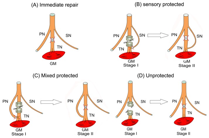
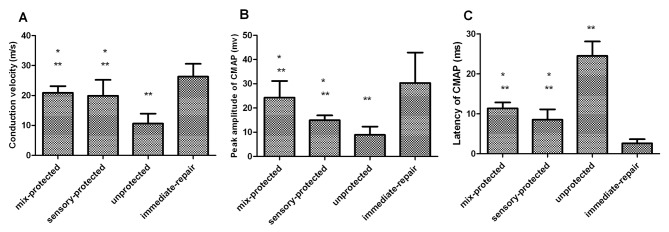
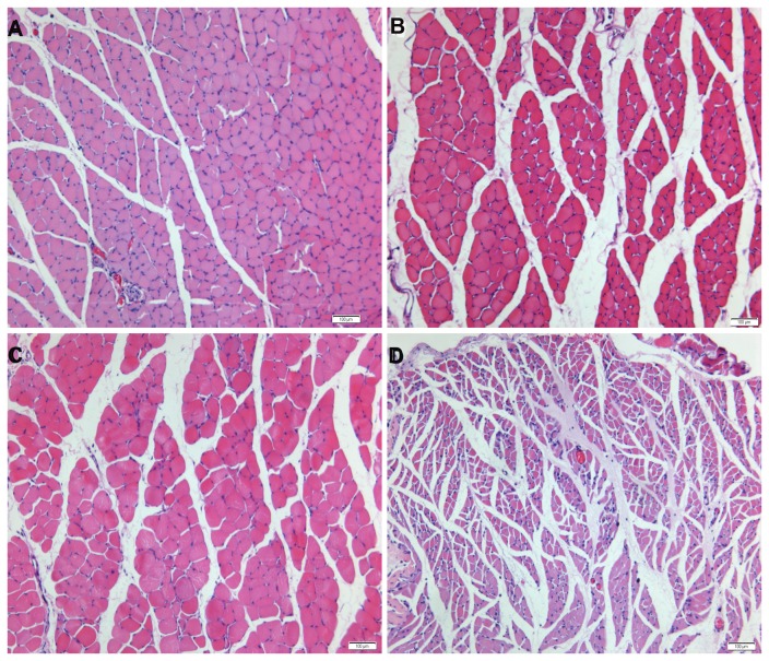
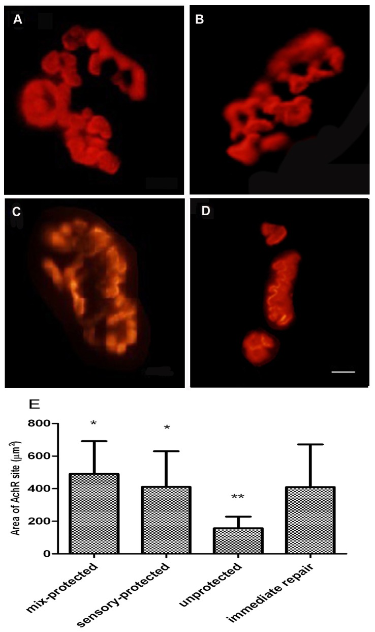
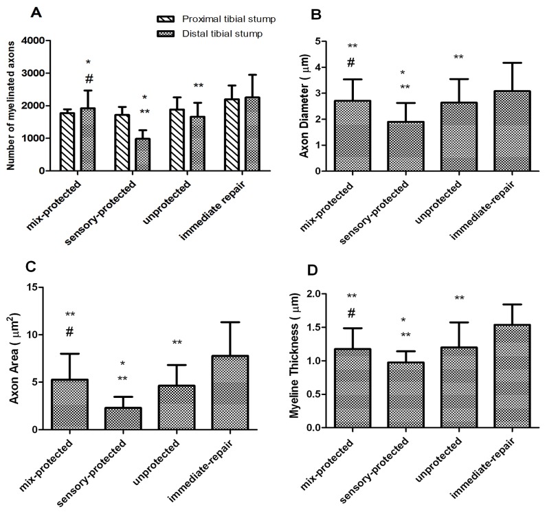
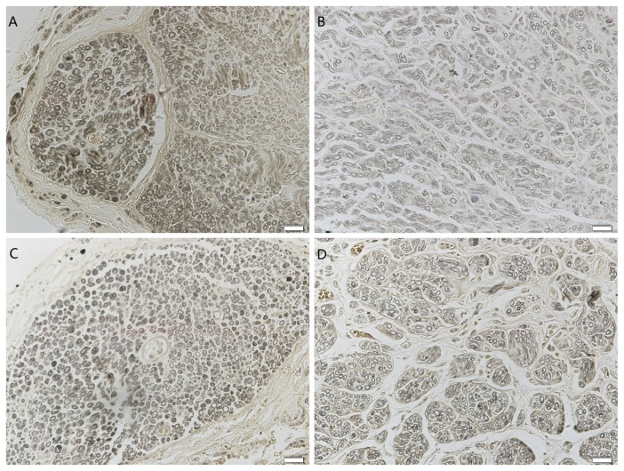
Functional recovery is usually poor following peripheral nerve injury when reinnervation is delayed. Early innervation by sensory nerve has been indicated to prevent atrophy of the denervated muscle. It is hypothesized that early protection with sensory axons is adequate to improve functional recovery of skeletal muscle following prolonged denervation of mixed nerve injury. In this study, four groups of rats received surgical denervation of the tibial nerve. The proximal and distal stumps of the tibial nerve were ligated in all animals except for those in the immediate repair group. The experimental groups underwent denervation with nerve protection of peroneal nerve (mixed protection) or sural nerve (sensory protection). The experimental and unprotected groups had a stage II surgery in which the trimmed proximal and distal tibial nerve stumps were sutured together. After 3 months of recovery, electrophysiological, histological and morphometric parameters were assessed. It was detected that the significant muscle atrophy and a good preserved structure of the muscle were observed in the unprotected and protective experimental groups, respectively. Significantly fewer numbers of regenerated myelinated axons were observed in the sensory-protected group. Enhanced recovery in the mixed protection group was indicated by the results of the muscle contraction force tests, regenerated myelinated fiber, and the results of the histological analysis. Our results suggest that early axons protection by mixed nerve may complement sensory axons which are required for promoting functional recovery of the denervated muscle natively innervated by mixed nerve.


Conflict of interest statement

Competing Interests: The authors have declared that no competing interests exist.

Figures

Figure 1
Figure 1. Schematic diagrams of surgical procedures producing chronic denervation of gastrocnemius muscle in the right hind limb of rats.
(A) Group 1- immediate tibial nerve repair. (B) Group 2- delayed tibial nerve repair with sensory nerve protection from the sural nerve in reverse end-to-side neurorrhaphy. (C) Group 3- delayed tibial nerve repair with mixed nerve protection from the peroneal nerve in reverse end-to-side neurorrhaphy. (D) Group 4- delayed tibial nerve repair without nerve protection. (TN: tibial nerve; PN: peroneal nerve; SN: sural nerve; GM: gastrocnemius muscle).
Figure 2
Figure 2. Results of electrophysiological examinations in the gastrocnemius muscle following denervation and subsequent repair with different manners.
The CMAP peak amplitudes, motor nerve conduction velocities and proximal latencies among the four groups were compared. There were significant differences between the unprotected group and all other groups and between immediate-repair group and all other groups, but there were no differences between mixed-protected group and sensory-protected group (*p < 0.05 compared with unprotected control; **p < 0.05 compared with immediate-repair control).
Figure 3
Figure 3. Photomicrographs of transverse sections of the gastrocnemius muscle following denervation and subsequent reinnervation.
The muscle sections of (A) immediate repair, (B) chronic denervation with sensory protection and (C) chronic denervation with mixed-nerve protection showed wide areas of larger muscle fibers among small groups of atrophied fibers. (D) Unprotected muscle sections showed extensive atrophy. Magnification×100. Scale bar= 100µm.
Figure 4
Figure 4. Postsynaptic AChR plaques at motor endplates in gastrocnemius muscle, as visualized by α-bungarotoxin binding.
(A) AChR plaques in the immediate repair group had well-defined contours. The appearance of AChR plaques in (B) chronic denervation with sensory protection and (C) chronic denervation with mixed-nerve protection displayed thick fringes and a few small round cupulae with distinct contours. The appearance of AChR plaques in (D) the unprotected group appeared as delineated, small, flat and slender. Magnification×400. Scale bar= 5µm. (E) Graphs showed that the area of AChR sites in the unprotected group was significantly smaller than in the other groups. (*p < 0.05 compared with unprotected control; **p < 0.05 compared with immediate-repair control).
Figure 5
Figure 5. Histograms depicting the assessment of the regenerated tibial nerve parameters.
(A) The number of myelinated axons were not significantly different among the four groups at the proximal region of the tibial nerve. At the distal transaction, the immediate-repair and mixed-protected rats had significantly greater number of regenerated myelinated axons than the unprotected ones. There were significantly fewer numbers of regenerated myelinated axons in the sensory-protected group. (B) The axonal diameter and (C) area of myelinated fibers were smaller and (D) the width of myelin sheath was thinner in sensory-protected group (*p < 0.05 compared with unprotected control; **p < 0.05 compared with immediate-repair control; # p < 0.05 compared with sensory-protected group).
Figure 6
Figure 6. Photomicrographs of the distal portion of the regenerated tibial nerve.
(A) Immediate repair showed good myelin regeneration. The diameters and thicknesses of the myelin sheathes were uniform and regular. (B) Chronic denervation with sensory protection displayed poor myelin regeneration, with an uneven distribution and a low density. A low level of myelin degeneration and necrosis was observed. The diameters and thicknesses in the sensory protection group were uneven and generally smaller than in the group of (C) chronic denervation with mixed-nerve protection. (D) The myelin distribution of delayed repair without protection was irregular. The diameters and thicknesses of the myelin sheathes in the unprotected group were generally smaller than in the mixed-protected group, and were larger than in the sensory-protected group. Magnification×400. Scale bar=20µm.

References

    1. Midha R (1997) Epidemiology of brachial plexus injuries in a multitrauma population. Neurosurgery 40: 1182-1189. - PubMed
    1. Gordon T, Tyreman N, Raji MA (2011) The basis for diminished functional recovery after delayed peripheral nerve repair. J Neurosci 31: 5325-5334. doi:10.1523/JNEUROSCI.6156-10.2011. PubMed: 21471367. - DOI - PMC - PubMed
    1. Höke A, Gordon T, Zochodne DW, Sulaiman OA (2002) A decline in glial cell-line-derived neurotrophic factor expression is associated with impaired regeneration after long-term Schwann cell denervation. Exp Neurol 173: 77-85. doi:10.1006/exnr.2001.7826. PubMed: 11771940. - DOI - PubMed
    1. Fu SY, Gordon T (1995) Contributing factors to poor functional recovery after delayed nerve repair: prolonged denervation. J Neurosci 15: 3886-3895. PubMed: 7751953. - PMC - PubMed
    1. Sulaiman OA, Gordon T (2000) Effects of short- and long-term Schwann cell denervation on peripheral nerve regeneration, myelination, and size. Glia 32: 234-246. doi:10.1002/1098-1136(200012)32:3. PubMed: 11102965. - DOI - PubMed

Publication types

Substances

LinkOut - more resources