. 2013 Nov 11;8(11):e79985.
doi: 10.1371/journal.pone.0079985. eCollection 2013.

Proton-sensing ovarian cancer G protein-coupled receptor 1 on dendritic cells is required for airway responses in a murine asthma model

Affiliations

Proton-sensing ovarian cancer G protein-coupled receptor 1 on dendritic cells is required for airway responses in a murine asthma model

Haruka Aoki et al. PLoS One. .

Abstract

Ovarian cancer G protein-coupled receptor 1 (OGR1) stimulation by extracellular protons causes the activation of G proteins and subsequent cellular functions. However, the physiological and pathophysiological roles of OGR1 in airway responses remain largely unknown. In the present study, we show that OGR1-deficient mice are resistant to the cardinal features of asthma, including airway eosinophilia, airway hyperresponsiveness (AHR), and goblet cell metaplasia, in association with a remarkable inhibition of Th2 cytokine and IgE production, in an ovalbumin (OVA)-induced asthma model. Intratracheal transfer to wild-type mice of OVA-primed bone marrow-derived dendritic cells (DCs) from OGR1-deficient mice developed lower AHR and eosinophilia after OVA inhalation compared with the transfer of those from wild-type mice. Migration of OVA-pulsed DCs to peribronchial lymph nodes was also inhibited by OGR1 deficiency in the adoption experiments. The presence of functional OGR1 in DCs was confirmed by the expression of OGR1 mRNA and the OGR1-sensitive Ca(2+) response. OVA-induced expression of CCR7, a mature DC chemokine receptor, and migration response to CCR7 ligands in an in vitro Transwell assay were attenuated by OGR1 deficiency. We conclude that OGR1 on DCs is critical for migration to draining lymph nodes, which, in turn, stimulates Th2 phenotype change and subsequent induction of airway inflammation and AHR.


Conflict of interest statement

Competing Interests: The authors have declared that no competing interests exist.

Figures

Figure 1
Figure 1. OGR1 mRNA expression in lung is increased by OVA sensitization/challenge.
WT and OGR1-/- mice were sensitized by OVA/alum and challenged by OVA. To nonsensitized mice, PBS was injected and inhaled. Lung mRNA expression of OGR1 and other proton-sensing GPCRs was measured 48 h after last antigen challenge. The results are mean + SEM of 9 determinations from three separate experiments. *The effect of OVA-priming (WT-PBS vs. WT-OVA) was significant. §The expression of OGR1 mRNA was undetectable.
Figure 2
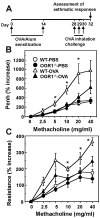
Figure 2. OGR1 deficiency attenuates AHR to methacholine in OVA-sensitized mice.
(A) OVA sensitization and challenge protocol. WT and OGR1 -/- mice were sensitized by i.p. injection of OVA/alum and challenged by OVA aerosol (triangle). To nonsensitized mice, PBS was injected and inhaled (circle). AHR to the inhaled methacholine was assessed by changes in noninvasive Penh (B) and dynamic resistance (C) 48 h after the last antigen exposure. Data are mean + SEM of n=13-16 per group. *p < 0.05 (WT-OVA vs. OGR1-/--OVA).
Figure 3
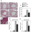
Figure 3. Histological analysis of lung sections.
Mice were sensitized and challenged by OVA or PBS as described in Figure 2. (A) Histological analysis of lung sections was performed 48 h after the last antigen exposure. The hematoxylin and PAS-stained sections shown are representative of three lung sections per mouse from 5 to 6 mice in each group. Scale bar, 200 μm. (B) The higher magnification image of the square in WT-OVA is shown. (C) Goblet cell hyperplasia was quantified as a PAS-staining cell number (C), and degree of peribronchial inflammation (D) and perivascular inflammation (E) was evaluated on a subjective scale of 0 to 3 as inflammatory score, in using the same sections as those used in (A). The column design specifying each group in (D) and (E) is the same as those in (C). Effects of OGR1 deficiency (WT-OVA vs. OGR1-/--OVA) were significant (*p < 0.05) throughout the figure.
Figure 4
Figure 4. OGR1 deficiency attenuates infiltration of inflammatory cells, Th2 cytokine levels in BAL fluids, and plasma IgE levels in OVA-sensitized mice.
Mice were sensitized and challenged by OVA or PBS as described in Figure 2. BAL fluid was analyzed 48 h after the last antigen exposure for total cell count and profile of cell types. Representative images of inflammatory cells (A) and differential cell counts (B) in BAL fluids. Arrowheads and arrows represent macrophages and eosinophils, respectively. Scale bar, 200 μm. Mac, macrophage; Lym, lymphocyte; Eos, eosinophils; and Neu, neutrophils. Data are mean + SEM of n=10-12 per group. The same BAL fluids were used to measure IL-4, IL-5, IL-13, and IFN-γ (C). Data are mean + SEM of n=10-12 per group. Plasma was collected for measurement of OVA-specific IgE levels after BAL fluid collection (D). Data are mean + SEM of n=6-15 per group. The column design specifying each group in (C) and (D) is the same as those in (B). The effects of OGR1 deficiency (WT-OVA vs. OGR1-/--OVA) were significant (*p < 0.05) throughout the figure.
Figure 5
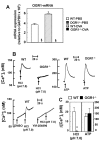
Figure 5. DCs express OGR1 mRNA and functional OGR1 activity.
DCs express OGR1 mRNA and functional OGR1 activity. Expression of OGR1 mRNA in DCs and its increase by OVA-priming (A). The expression of OGR1 mRNA was evaluated by quantitative real-time TaqMan PCR. Data are mean ± SEM of 9 determinations from three separate experiments. Effect of OVA-priming (WT-PBS vs. WT-OVA) was significant (*p < 0.05). §The expression of OGR1 mRNA was undetectable. Acidification induces [Ca2+]i increase through the OGR1/Gq/11-protein in DCs (B and C). DCs were prepared from WT and OGR1 -/- mice and incubated to monitor [Ca2+]i evaluated by the change in Fura-2 fluorescence. The cells were preincubated in suspension at pH 7.4 and, at arrow, HCl (final pH of 7.0) or ATP (100 μM) was added. To evaluate the role of Gq/11-protein, the cell suspension was treated with YM-254890 (100 nM) (or DMSO as vehicle). A representative trace of [Ca2+]i change was shown (B). Net [Ca2+]i change (peak value - basal value) was evaluated (C). Basal value of [Ca2+]i at pH 7.4 was 125 ± 12 nM and the value was not appreciably affected by OGR1 deficiency. Data are mean ± SEM of 8 determinations from four separate experiments. Effect of OGR1 deficiency (WT vs. OGR1-/-) was significant (*p < 0.05).
Figure 6
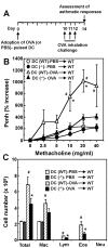
Figure 6. Intratracheal administration of OGR1-deficient DCs showed the lowered AHR and eosinophilia compared with that of WT DCs.
(A) Protocol of DC adoption experiment. OVA or PBS-pulsed BMDCs were prepared from WT and OGR1 -/- mice and administered intratracheally to WT mice. Ten days later, mice were challenged with OVA inhalation for 20 min on three consecutive days. Forty-eight h after the last OVA-challenge, AHR to methacholine was measured as Penh activities (B). After measurement of airway responsiveness, BAL fluids were collected and analyzed for the count of total cells and differentiated cells (C). Data are mean + SEM of n=10-12 per group. # p < 0.05 (DC (WT)-PBS vs. DC (WT)-OVA) and *p < 0.05 (DC (WT)-OVA vs. DC (-/-)-OVA).
Figure 7
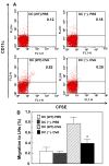
Figure 7. DC migration to peribronchial lymph nodes is inhibited by OGR1 deficiency.
Four groups of CFSE-labeled DCs, i.e., PBS-treated WT DC, PBS-treated OGR1-deficient DC, OVA-treated WT DC, and OVA-treated OGR1-deficient DC, were instilled intratracheally to naïve WT mice and the CFSE+ DCs migrating in the peribronchial lymph nodes 96 h later were analyzed by flow cytometry. (A) Cells recovered from lymph nodes were assessed for the expression of CD11c and CFSE. Representative flow cytometry plots from 3 separate experiments per group are shown. (B) Percentages of CFSE+DCs (CFSE+CD11c+) per total cells applied were shown. Data are mean ± SEM of 6 analyses from 3 separate experiments. Effect of OGR1 deficiency (DC (WT)-OVA vs. DC (-/-)-OVA) was significant (*p < 0.05).
Figure 8
Figure 8. Mature DC migration is attenuated in association with reduced expression of CCR7 by OGR1 deficiency in vitro.
(A and B) BMDCs from WT or OGR1-deficient mice were pulsed with 100 μg/ml of OVA as mature DCs or its vehicle as immature DCs overnight. Migration response to the indicated concentrations of ATP for non-pulsed immature DCs (A) and to 200 ng/ml CCL19 or CCL21 for OVA-pulsed DCs (B) was measured. Results are expressed as percentages of basal values obtained without any agonist in DCs of WT mice. Data are mean ± SEM of 5 separate experiments. (C) The expression of CCR7 protein was analysed by flow cytometry. Representative results of four separate experiments in each group are shown. CCR7-protein expression is also expressed as mean fluorescence intensity (MFI) + SEM. (D) The expression of CCR7 mRNA was measured by quantitative real-time TaqMan PCR. Data are mean ± SEM of 9 determinations from three separate experiments. The effects of OGR1 deficiency (WT-OVA vs. OGR1-/--OVA) were significant throughout the figure (*p < 0.05).

References

    1. Lambrecht BN, Hammad H (2009) Biology of lung dendritic cells at the origin of asthma. Immunity 31: 412-424. doi:10.1016/j.immuni.2009.08.008. PubMed: 19766084. - DOI - PubMed
    1. Holgate ST (2012) Innate and adaptive immune responses in asthma. Nat Med 18: 673-683. doi:10.1038/nm.2731. PubMed: 22561831. - DOI - PubMed
    1. Hunt JF, Fang K, Malik R, Snyder A, Malhotra N et al. (2000) Endogenous airway acidification. Implications for asthma pathophysiology. Am J Respir Crit Care Med 161: 694-699. doi:10.1164/ajrccm.161.3.9911005. PubMed: 10712309. - DOI - PubMed
    1. Ricciardolo FL, Gaston B, Hunt J (2004) Acid stress in the pathology of asthma. J Allergy Clin Immunol 113: 610-619. doi:10.1016/j.jaci.2003.12.034. PubMed: 15100663. - DOI - PubMed
    1. Kodric M, Shah AN, Fabbri LM, Confalonieri M (2007) An investigation of airway acidification in asthma using induced sputum: a study of feasibility and correlation. Am J Respir Crit Care Med 175: 905-910. doi:10.1164/rccm.200607-940OC. PubMed: 17290044. - DOI - PubMed

Publication types

MeSH terms