. 2013 Nov 14;8(11):e80104.
doi: 10.1371/journal.pone.0080104. eCollection 2013.

Hearing loss in a mouse model of 22q11.2 Deletion Syndrome

Affiliations

Hearing loss in a mouse model of 22q11.2 Deletion Syndrome

Jennifer C Fuchs et al. PLoS One. .

Abstract

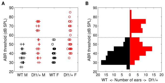
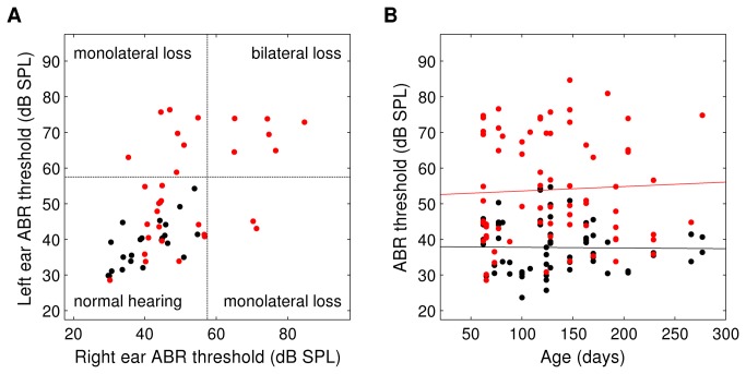
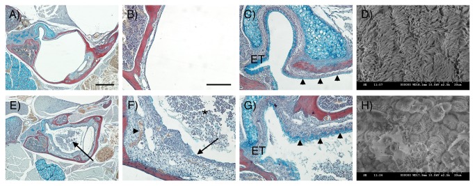
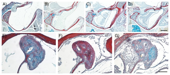
22q11.2 Deletion Syndrome (22q11DS) arises from an interstitial chromosomal microdeletion encompassing at least 30 genes. This disorder is one of the most significant known cytogenetic risk factors for schizophrenia, and can also cause heart abnormalities, cognitive deficits, hearing difficulties, and a variety of other medical problems. The Df1/+ hemizygous knockout mouse, a model for human 22q11DS, recapitulates many of the deficits observed in the human syndrome including heart defects, impaired memory, and abnormal auditory sensorimotor gating. Here we show that Df1/+ mice, like human 22q11DS patients, have substantial rates of hearing loss arising from chronic middle ear infection. Auditory brainstem response (ABR) measurements revealed significant elevation of click-response thresholds in 48% of Df1/+ mice, often in only one ear. Anatomical and histological analysis of the middle ear demonstrated no gross structural abnormalities, but frequent signs of otitis media (OM, chronic inflammation of the middle ear), including excessive effusion and thickened mucosa. In mice for which both in vivo ABR thresholds and post mortem middle-ear histology were obtained, the severity of signs of OM correlated directly with the level of hearing impairment. These results suggest that abnormal auditory sensorimotor gating previously reported in mouse models of 22q11DS could arise from abnormalities in auditory processing. Furthermore, the findings indicate that Df1/+ mice are an excellent model for increased risk of OM in human 22q11DS patients. Given the frequently monaural nature of OM in Df1/+ mice, these animals could also be a powerful tool for investigating the interplay between genetic and environmental causes of OM.


Conflict of interest statement

Competing Interests: The authors have declared that no competing interests exist.

Figures

Figure 1
Figure 1. Elevated ABR thresholds, and bimodal distribution of thresholds, in Df1/+ mice.
(A) Click ABR thresholds recorded from individual ears in male and female WT (black) and Df1/+ (red) mice. Median ABR thresholds differed significantly between Df1/+ and WT mice of the same gender (Wilcoxon Mann-Whitney test, Df1/+ versus WT: p=6x10⁻⁶ males, p=9x10⁻⁷ females), but not between males and females of the same genotype (Wilcoxon Mann-Whitney test, males versus females: p=0.3 Df1/+, p=0.5 WT). (B) Click ABR thresholds pooled across recordings from male and female animals, to illustrate the bimodal appearance of the Df1/+ ABR threshold distribution. Dashed lines indicate the criterion threshold for a significant click ABR deficit (see text).
Figure 2
Figure 2. ABR deficit in Df1/+ mice is often monolateral, and shows no age dependence.
(A) Click ABR thresholds recorded in left versus right ears, for WT (black) and Df1/+ (red) mice in which both ears were tested. Dashed line indicates criterion threshold for a significant click ABR deficit. (B) Click ABR thresholds versus age, for all ABR recordings. Solid lines indicate best-fit regression lines. Slopes were not significantly different from zero for recordings from either WT (black; slope 95% CI [-0.047, 0.072]) or Df1/+ (red; slope 95% CI [-0.072, 0.030]) animals. To ensure visibility of overlapped data points, zero-mean, 1 dB SPL standard-deviation Gaussian noise was added to threshold data in both A and B for display.
Figure 3
Figure 3. Morphological changes in MEC mucosa and epithelium in Df1/+ mice.
(A-C, E-G) Frontal trichrome-stained sections from 11.5-week-old mice showing the middle ear cavity. (D, H) SEM images of middle ear epithelium. (A-D) WT. (E-H) Df1/+ with signs of OM. (A) The middle ear cavity is air-filled in the WT. (B) The mucosa is a thin layer lining the auditory bulla. (C) At the entrance of the Eustachian tube (ET) high levels of alcian blue staining are observed indicating mucin production. Further into the middle ear away from the orifice, staining is less distinct in the WT (arrowheads). (D) A thick lawn of cilia is observed overlying the epithelium near the ET orifice. (E) In Df1/+ mice the middle ear cavity is filled with effusion (arrow). (F) Df1/+ mice show signs of inflammation such as effusion with infiltrated inflammatory cells (asterix), a thickened mucosa (arrow) and hypervascularisation (arrowhead). (G) In addition, increased alcian blue staining is observed within the middle ear at a distance from the ET indicating increased mucin production in Df1/+ mice (compare C and G, arrowheads). (H) Df1/+ mice with OM show reduced numbers of cilia that appear shortened and rarefied. Dorsal is top in A-C, E-G. Scale bar: 500 μm (A, E), 100 μm (B, C, F, G).
Figure 4
Figure 4. The severity of OM correlates with the degree of hearing loss in Df1/+ mice (see also Table 1).
(A-G) Frontal trichrome-stained sections from adult mice showing the graded severity of effusion in Df1/+ middle ear cavities (A-D) and thickened mucosa around the head of the malleus (E-G). Least severe conditions are displayed on the left hand side panels with increasing severity towards the right hand side panels. (A) No effusion, (B) serous effusion, (C) effusion with <50% infiltrated cells and (D) effusion with >50% infiltrated cells. (E) No thickening, (F) mild thickening and (G) severe thickening of the mucosa around the head of the malleus. Dorsal is top. Scale bar: 500 μm (A-D), 100 μm (E-G).

References

    1. Scambler PJ (2000) The 22q11 deletion syndromes. Hum Mol Genet 9: 2421-2426. doi:10.1093/hmg/9.16.2421. PubMed: 11005797. - DOI - PubMed
    1. Scambler PJ (2010) 22q11 deletion syndrome: a role for TBX1 in pharyngeal and cardiovascular development. Pediatr Cardiol 31: 378-390. doi:10.1007/s00246-009-9613-0. PubMed: 20054531. - DOI - PubMed
    1. Paylor R, Lindsay E (2006) Mouse models of 22q11 deletion syndrome. Biol Psychiatry 59: 1172-1179. doi:10.1016/j.biopsych.2006.01.018. PubMed: 16616724. - DOI - PubMed
    1. Lindsay EA, Baldini A (1998) Congenital heart defects and 22q11 deletions: which genes count? Mol Med Today 4: 350-357. doi:10.1016/S1357-4310(98)01302-1. PubMed: 9755454. - DOI - PubMed
    1. Ryan AK, Goodship JA, Wilson DI, Philip N, Levy A et al. (1997) Spectrum of clinical features associated with interstitial chromosome 22q11 deletions: a European collaborative study. J Med Genet 34: 798-804. doi:10.1136/jmg.34.10.798. PubMed: 9350810. - DOI - PMC - PubMed

Publication types

MeSH terms

LinkOut - more resources