. 2013 Nov 14;8(11):e80181.
doi: 10.1371/journal.pone.0080181. eCollection 2013.

Expression and secretion of plasma membrane Ca2+-ATPase 4a (PMCA4a) during murine estrus: association with oviductal exosomes and uptake in sperm

Affiliations

Expression and secretion of plasma membrane Ca2+-ATPase 4a (PMCA4a) during murine estrus: association with oviductal exosomes and uptake in sperm

Amal A Al-Dossary et al. PLoS One. .

Abstract

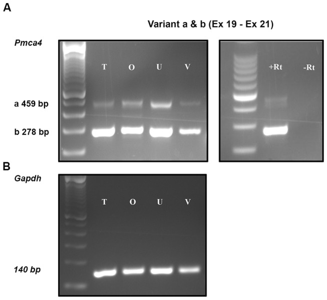
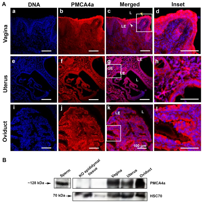
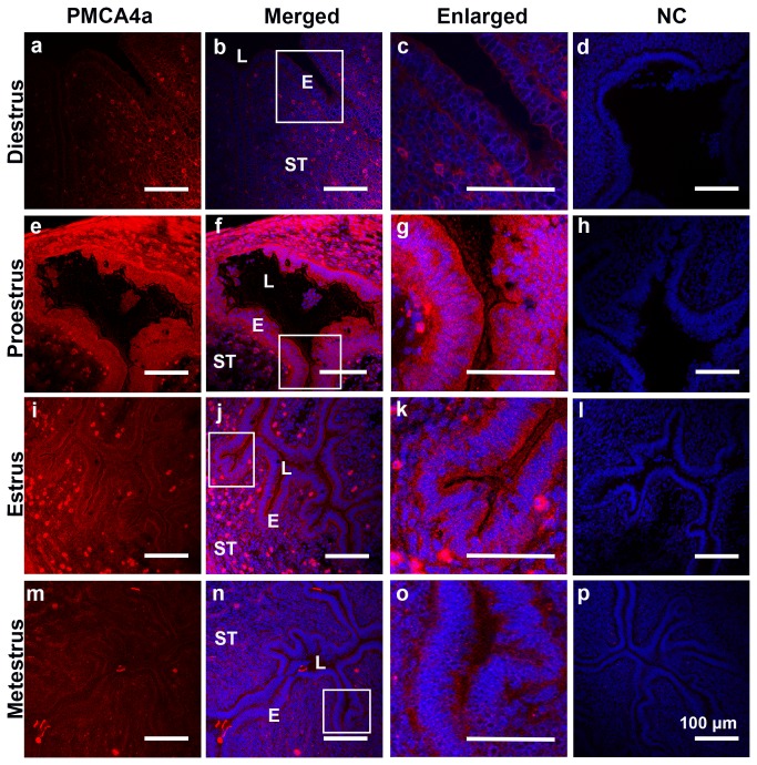
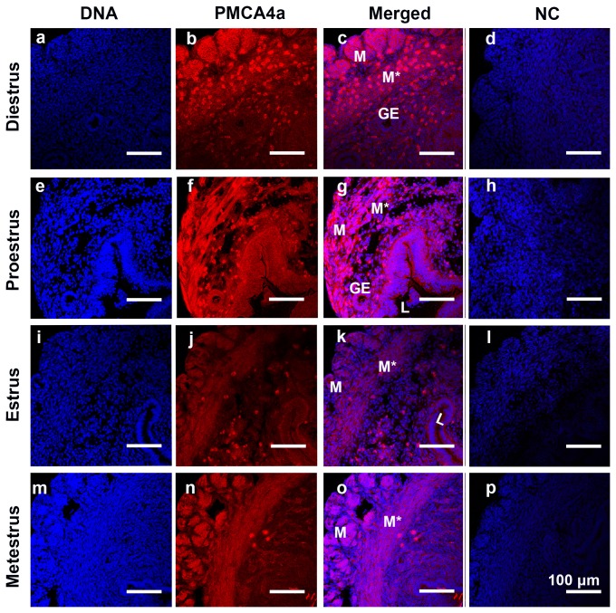
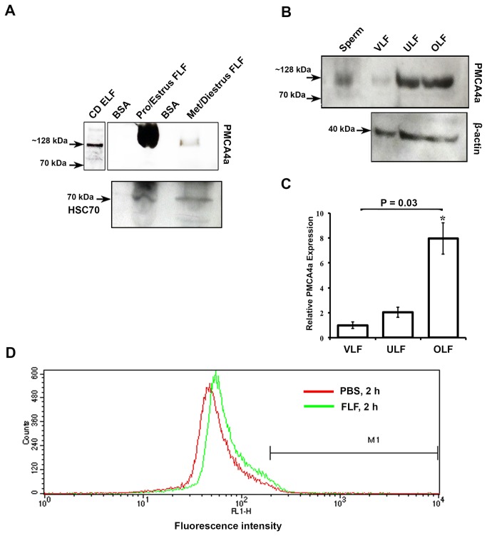
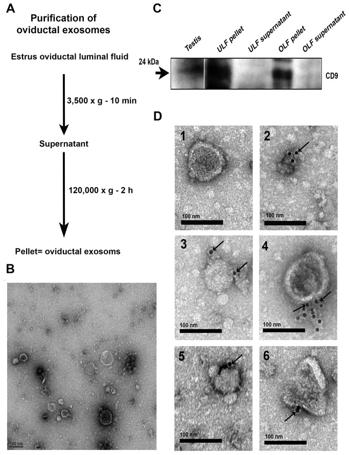
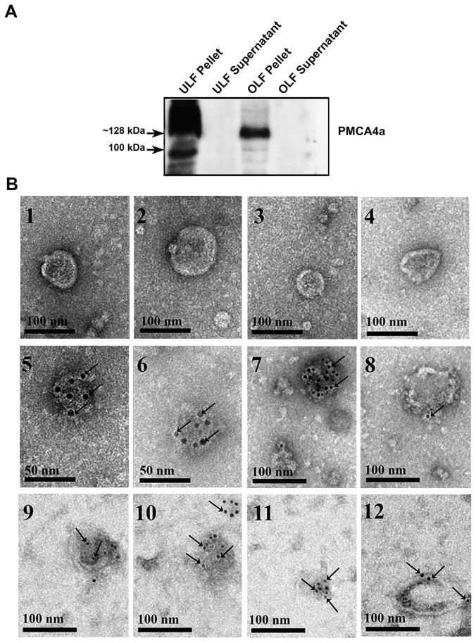
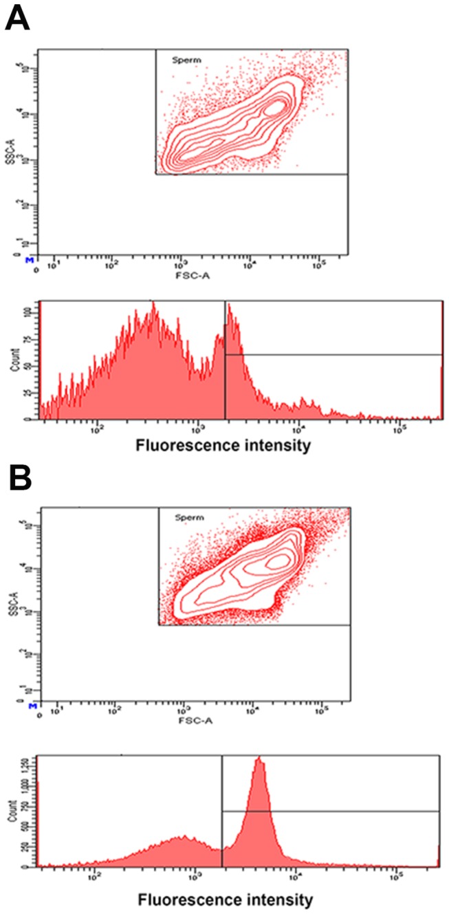
PMCA4, a membrane protein, is the major Ca(2+) efflux pump in murine sperm where its deletion leads to a severe loss of hyperactivated motility and to male infertility. We have previously shown that the PMCA4b splice variant interacts with CASK (Ca(2+/)CaM-dependent serine kinase) in regulating sperm Ca(2+). More recently we detected that PMCA4a isoform, in addition to its presence in testis, is secreted in the epididymal luminal fluid and transferred to sperm. Here we show that Pmca4 mRNA is expressed in both the 4a and 4b variants in the vagina, uterus, and oviduct. Immunofluorescence reveals that PMCA4a is similarly expressed and is elevated during estrus, appearing in the glandular and luminal epithelia. Western analysis detected PMCA4a in all tissues and in the luminal fluids (LF) of the vagina (VLF), uterus (ULF), and the oviduct (OLF) collected during estrus. It was ~9- and 4-fold higher in OLF than in VLF and ULF, and only marginally present in LF collected at metestrus/diestrus. Fractionation of the LF collected at estrus, via ultracentrifugation, revealed that 100% of the PMCA4a resides in the vesicular fraction of the ULF and OLF. Transmission electron microscopy (TEM) revealed that OLF vesicles have an exosomal orientation (with the cytoplasmic-side inward), a size range of 25-100 nm, with the characteristic CD9 biomarker. Thus, we dubbed these vesicles "oviductosomes", to which PMCA4a was immunolocalized. Incubation of caudal sperm in the combined LF or exosomes resulted in up to a ~3-fold increase of sperm PMCA4a, as detected by flow cytometry, indicating in vitro uptake. Our results are consistent with the increased requirement of Ca(2+) efflux in the oviduct. They show for the first time the presence of oviductal exosomes and highlight their role, along with uterosomes and vaginal exosomes, in post-testicular sperm acquisition of PMCA4a which is essential for hyperactivated motility and fertility.


Conflict of interest statement

Competing Interests: The authors have declared that no competing interests exist.

Figures

Figure 1
Figure 1. Semi-quantitative analysis of Pmca4a, b mRNA via RT-PCR in the murine female reproductive tract
A) The 469 bp PCR product represents Pmca4a, and that of 278 bp corresponds to Pmca4b. Murine testis (T) cDNA served as positive control. Negative control was performed in the absence of reverse transcriptase (-RT). Gapdh mRNA was used as an internal control (B). Pronounced expression of Pmca4b was detected in the uterus, whereas PMCA4a showed more prominent expression in the oviduct than in the testis. A 100 bp ladder was run in the left lane of each panel. O, oviduct; U, uterus, and V, vagina.
Figure 2
Figure 2. Expression of PMCA4a in mouse vaginal, uterine, and oviductal tissues following superovulation
A) Indirect immunofluorescence was performed on frozen sections (a-d) of vaginal tissues using anti-PMCA4a antibody and an AlexaFluor-conjugated secondary antibody (red), and the nuclei were visualized by staining with Draq-5 (blue). Strong PMCA4a staining was detected at the epithelial layers at the luminal edge (yellow arrowhead) and was decreased at the basement membrane (white arrowhead). (e-h) In uterine tissue PMCA4a was abundantly expressed in both the luminal and glandular epithelia and also in the stroma. (i-l) In oviductal tissue strong PMCA4a staining is detected at the apical boundaries of the epithelial cells that line the oviductal lumen. (d, h, and l) Insets are seen from vagina, uterus and oviduct. Negative controls in PBS or IgG of the tissues showed no staining, similar to that in Figure 3, 4. The images were captured using confocal microscopy with a 20x objective lens (a plan-Apochromatic). L= lumen; LE = luminal epithelium; GE = glandular epithelium. Bar = 100 µm (same scale for all micrographs, and 200 µm insets). B) Western blot analysis performed with anti-PMCA4a antibody on tissues recovered after superovulation, using sperm as a positive control, revealed the ~128 kDa PMCA4a in all tissues and its absence in epididymal tissues of Pmca4 null mice, used as a negative control (Top panel). In the lower panel, equal loading of protein is demonstrated by detection of HSC 70 in the tissue lysates. The amount of proteins loaded was 20 μg per lane.
Figure 3
Figure 3. Indirect Immunofluorescence of PMCA4a in the murine endometrium of the uterus during the estrous cycle
Using frozen sections PMCA4a immunoreactivity (red) was detected in the uterine luminal epithelium in pro-estrus (e - g) and estrus phases (i - k) but not during metestrus (m - o) and diestrus (a - c). The nuclei were visualized by staining with Draq-5 (blue). Negative controls (NC) in PBS or IgG are shown in d, h, l, and p. The images were captured using confocal microscopy and a 20x (a plan-Apochromatic) objective lens. LE = luminal epithelium; L= lumen; ST= stroma. Bar = 100 µm (same scale for all micrographs, and 200 µm for insets).
Figure 4
Figure 4. Indirect Immunofluorescence of PMCA4a in the murine myometrium of the uterus during the estrous cycle
In addition to the endometrium, the muscles and mesothelium of the myometrium (M*, M, respectively) were positively stained for PMCA4a. Elevated levels of PMCA4a immunoreactivity were detected at the boundaries of the epithelial cells lining the pro-estrus uterine glands (g). The nuclei were visualized by staining with Draq-5 (blue). Negative controls (NC) of diestrus, pro-estrus, estrus, and metestrus phases are respectively shown (d, h, l, and p). The images were captured using confocal microscopy and a 20x (a plan-Apochromatic) objective lens. GE = glandular epithelium; L = lumen; ST= stroma. Bar = 100 µm (same scale for all micrographs).
Figure 5
Figure 5. Detection of PMCA4a in reproductive luminal fluids and its acquisition on caudal sperm
A) Representative Western blot of FLFs collected during pro-estrus and estrus and metestrus and diestrus (40 µg proteins loaded). The ~128 kDa PMCA4a is seen in pro-estrus and estrus and is marginally present at metestrus and diestrus. Caudal epididymal luminal fluid was used as a positive control. The membrane was stripped and re-probed for HSC70 as a loading control. B) Western blots of VLF, ULF, and OLF recovered after superovulation demonstrate the presence of the ~128 kDa PMCA4a. Sperm protein was used as a positive control. The membrane was stripped and re-probed for β-actin as a loading control. C) Quantitation of Western blot data shown in B; the relative expression was determined using VLF as 1. The data represent the mean (±SEM) of a minimum of three independent experiments, and the intensity was quantified by Image J software. ANOVA and t-tests were performed on the mean and P values were calculated. *P = 0.03 indicates a significantly increase amount of PMCA4a in OLF compared to that in VLF. D) A peak shift of fluorescence intensity to the right, indicates increase amounts of PMCA4a in sperm incubated in FLF compared to PBS for 2 h and treated as described in Materials and Methods.
Figure 6
Figure 6. Characterization of membranous vesicles in OLF.
A) Protocol used to isolate oviductal exosomes by ultracentrifugation of oviductal fluids. B) TEM of negative staining for the particulate fraction from OLF reveals the presence of membranous vesicles ranging in size from 25-100 nm in diameter. C) Western blots detected CD9 (24 kDa) in protein extracts from membranous vesicles removed from OLF and uterosomes, but not in the supernatants. Testis protein was used as a positive control. Each lane contains 40 µg of protein. Results are representative of three different experiments. D) Immunogold labeling (6 nm gold particles) of CD9 is shown in oviductal membranous vesicles termed “oviductosomes”. Gold particles on individual oviductosomes are seen arrowed in 2-6 on the exterior of the membrane. In the absence of primary antibodies and the presence of rat IgG, gold particles were absent (1), indicating the specificity of the antibody. Scale bar =100 nm in panel B, D.
Figure 7
Figure 7. Immunodetection of PMCA4a in oviductosomes and uterosomes.
A) Nitrocellulose membrane with CD-positive pellets and supernatants (Figure 6C), when stripped and re-probed with PMCA4a antibodies in a Western blot, revealed the presence of the ~128 kDa PMCA4a band in the pellets only. A band of unknown origin at ~100 kDa is also seen in the uterosomes. Each lane contains 40 µg of proteins. Results are representative of three experiments. B) Immunogold labeling (6 nm gold particles) of PMCA4a is shown in oviductosomes in 5-8 and in uterosomes in 9-12. Gold particles localized on the cytoplasmic-side of the membrane are seen arrowed. They were rarely seen elsewhere on the grids. In the absence of primary antibodies and the presence of rabbit IgG, gold particles were absent (1-4) indicating the specificity of the antibody. Scale bar= 50-100 nm.
Figure 8
Figure 8. PMCA4a uptake by caudal sperm via incubation in exosomes reconstituted in PBS
Exosomes were isolated from FLFs after superovulation. A) Flow cytometric analysis of sperm co-incubated in PBS (negative control). The contour plot above reveals two subpopulations of sperm, as represented by the small inner circles at top right and bottom left. The graph shows that the majority of the fluorescence falls below the gated region. B) Sperm incubated in FLF exosomes (2 mg/ml protein) show a unimodal distribution (a single inner circle) and a ~3 fold increase of fluorescence intensity (peak shift to the right) compared to A in the gated region. The co-incubation period was 3 h for both A and B.

References

    1. Rodriguez-Martinez H (2007) Role of the oviduct in sperm capacitation. Theriogenology 68 Suppl 1: S138-S146. doi:10.1016/j.theriogenology.2007.03.018. PubMed: 17452049. - DOI - PubMed
    1. de Lamirande E, Leclerc P, Gagnon C (1997) Capacitation as a regulatory event that primes spermatozoa for the acrosome reaction and fertilization. Mol Hum Reprod 3: 175-194. doi:10.1093/molehr/3.3.175. PubMed: 9237244. - DOI - PubMed
    1. Wennemuth G, Babcock DF, Hille B (2003) Calcium clearance mechanisms of mouse sperm. J Gen Physiol 122: 115-128. doi:10.1085/jgp.200308839. PubMed: 12835474. - DOI - PMC - PubMed
    1. Okunade GW, Miller ML, Pyne GJ, Sutliff RL, O'Connor KT et al. (2004) Targeted ablation of plasma membrane Ca2+-ATPase (PMCA) 1 and 4 indicates a major housekeeping function for PMCA1 and a critical role in hyperactivated sperm motility and male fertility for PMCA4. J Biol Chem 279: 33742-33750. doi:10.1074/jbc.M404628200. PubMed: 15178683. - DOI - PubMed
    1. Schuh K, Cartwright EJ, Jankevics E, Bundschu K, Liebermann J et al. (2004) Plasma membrane Ca2+ ATPase 4 is required for sperm motility and male fertility. J Biol Chem 279: 28220-28226. doi:10.1074/jbc.M312599200. PubMed: 15078889. - DOI - PubMed

Publication types

MeSH terms

LinkOut - more resources