. 2013 Nov 7;8(11):e80695.
doi: 10.1371/journal.pone.0080695. eCollection 2013.

MuSK myasthenia gravis IgG4 disrupts the interaction of LRP4 with MuSK but both IgG4 and IgG1-3 can disperse preformed agrin-independent AChR clusters

Affiliations

MuSK myasthenia gravis IgG4 disrupts the interaction of LRP4 with MuSK but both IgG4 and IgG1-3 can disperse preformed agrin-independent AChR clusters

Inga Koneczny et al. PLoS One. .

Abstract

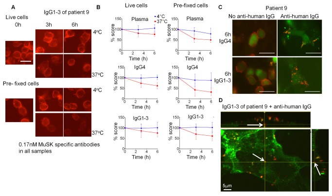
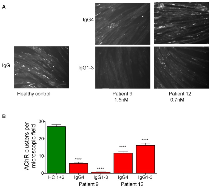
A variable proportion of patients with generalized myasthenia gravis (MG) have autoantibodies to muscle specific tyrosine kinase (MuSK). During development agrin, released from the motor nerve, interacts with low density lipoprotein receptor-related protein-4 (LRP4), which then binds to MuSK; MuSK interaction with the intracellular protein Dok7 results in clustering of the acetylcholine receptors (AChRs) on the postsynaptic membrane. In mature muscle, MuSK helps maintain the high density of AChRs at the neuromuscular junction. MuSK antibodies are mainly IgG4 subclass, which does not activate complement and can be monovalent, thus it is not clear how the antibodies cause disruption of AChR numbers or function to cause MG. We hypothesised that MuSK antibodies either reduce surface MuSK expression and/or inhibit the interaction with LRP4. We prepared MuSK IgG, monovalent Fab fragments, IgG1-3 and IgG4 fractions from MuSK-MG plasmas. We asked whether the antibodies caused endocytosis of MuSK in MuSK-transfected cells or if they inhibited binding of LRP4 to MuSK in co-immunoprecipitation experiments. In parallel, we investigated their ability to reduce AChR clusters in C2C12 myotubes induced by a) agrin, reflecting neuromuscular development, and b) by Dok7- overexpression, producing AChR clusters that more closely resemble the adult neuromuscular synapse. Total IgG, IgG4 or IgG1-3 MuSK antibodies were not endocytosed unless cross-linked by divalent anti-human IgG. MuSK IgG, Fab fragments and IgG4 inhibited the binding of LRP4 to MuSK and reduced agrin-induced AChR clustering in C2C12 cells. By contrast, IgG1-3 antibodies did not inhibit LRP4-MuSK binding but, surprisingly, did inhibit agrin-induced clustering. Moreover, both IgG4 and IgG1-3 preparations dispersed agrin-independent AChR clusters in Dok7-overexpressing C2C12 cells. Thus interference by IgG4 antibodies of the LRP4-MuSK interaction will be one pathogenic mechanism of MuSK antibodies, but IgG1-3 MuSK antibodies will also contribute to the reduced AChR density and neuromuscular dysfunction in myasthenia patients with MuSK antibodies.


Conflict of interest statement

Competing Interests: The authors have read the journal's policy and have the following conflicts. The University of Oxford holds the following patent for MuSK antibody for diagnosis of myasthenia (with the late Dr. W. Hoch, University of Tuebingen) PCT/GB01/02661. Licensed to Athena Diagnostics USA who sublicensed to RSR Ltd in 2003; royalties to Oxford University. AV is a named inventor and receives a share of royalties. AV also receives consulting fees (5K pa) from Athena Diagnostics. This does not alter the authors' adherence to all the PLOS ONE policies on sharing data and materials. The other authors have declared that no competing interests exist.

Figures

Figure 1
Figure 1. Characterisation of antibodies from MuSK-MG patients.
(A) MuSK antibody titres of 14 MuSK MG patients, 30 healthy controls and 5 AChR MG patients as determined by RIA. Cut-off for MuSK antibody positivity was the mean titre of pooled healthy controls + 3x standard deviation. (B) Example of IgG subclass profiles from 3 MuSK patients. Patient plasma was incubated with HEK293 cells expressing MuSK, or to mock-transfected HEK293 cells. A subclass-specific secondary antibody was added, followed by a fluorescent tertiary antibody, and the amount of binding (mean fluorescence intensity, mfi) was measured by flow cytometry. Δmfi is the mfi obtained with cells expressing MuSK minus the mfi with mock-transfected cells. (C) Correlation between IgG subclasses, measured by flow cytometry as for (B), and MuSK antibody titres measured by RIA of 14 MuSK patient plasmas. Statistic analysis: linear regression followed by Pearson correlation (Gaussian distribution was determined by D’Agostino and Pearson test). (D) IgG1-3 and IgG4 subclasses were purified from MuSK-MG patients 9 and 12. The purity of each of these two subclass groups was analysed by flow cytometry as for (B). The IgG4 fraction for both patients contains some contamination of IgG2, but the IgG1-3 fraction is devoid of IgG4 for both patients.
Figure 2
Figure 2. MuSK patient IgG4 or IgG1-3 do not induce endocytosis of MuSK.
Patient plasma or purified IgG1-3 or IgG4 were applied at 0.17nM final concentration of MuSK antibody to HEK293 cells expressing MuSK at the cell surface. Cells were either incubated at 4°C to prevent, or at 37°C to allow, endocytosis. Patient antibody binding was visualised by addition of a secondary fluorescent anti-human antibody at the end of the experiment. (A) An example of cells that were incubated with IgG1-3 from patient 9. Robust staining was observed after 6 hours incubation at both temperatures. Scale bar=25µm. (B) Staining was scored by two individuals as described in methods, and the scores were normalised to the score at 0 hours for each condition. A slight decrease in staining was observed for cells incubated at 37°C, and pre-fixed cells also showed this reduction. (C) As a positive control for endocytosis, IgG1-3 or IgG4 was cross-linked by addition of Alexa Fluor 568-conjugated anti-human IgG prior to incubation. Example images show cells treated with patient 9 IgG4 and IgG1-3 in the presence and absence of cross-linking anti-human IgG after 6 hours incubation at 37°C. There is a clear difference when the cross-linking secondary antibody is present. Scale bar =50µm (D) Cross-linking by the secondary antibody induced internalisation of human anti-MuSK antibodies as well as MuSK-EGFP as early as 30 minutes, as observed by confocal microscopy. An example image of a cell from a confocal z-stack with orthogonal side-views is shown. The arrow shows internalised secondary Alexa Fluor 568-conjugated anti-human IgG (red) colocalised with EGFP-tagged MuSK (green). Scale bar =5µm.
Figure 3
Figure 3. Both IgG4 and IgG1-3 subclass antibodies impair agrin-induced AChR clustering in C2C12 myotubes.
C2C12 myotubes were incubated overnight with plasma from a healthy control, or with purified IgG4 or IgG1-3 at final concentrations of 0.7nM MuSK antibodies (patient 12) or 1.5nM (patient 9), in the presence of agrin. AChR clusters were stained with Alexa Fluor 594-conjugated α−bungarotoxin and 30 microscopic images at 20x magnifications were acquired. (A) Example images showing AChR clusters, scale bar =50µm. (B) Quantification of AChR clusters per field using ImageJ software (N=3). Statistical analysis: one way ANOVA (p=0.0001) followed by Bonferroni post test. **** p≤0.0001.
Figure 4
Figure 4. MuSK antibodies block binding between MuSK and LRP4.
HEK293 cells expressing MuSK-mCherry and/or LRP4-EGFP were used to study MuSK-LRP4 binding using co-immunoprecipitation (co-IP). (A, B) Demonstration that MuSK and LRP4 interact. (A) An EGFP-specific antibody was able to co-immunoprecipitate LRP4-EGFP and MuSK-mCherry (detected with an mCherry-specific antibody) only from the surface of cells that were transfected with both LRP4-EGFP and MuSK-mCherry. (B) The MuSK-specific AF562 antibody could co-immunoprecipitate MuSK-mCherry and LRP4-EGFP only from the surface of cells transfected with both proteins. Healthy control serum (HC) did not influence this co-precipitation. (C) MuSK-MG patient plasma was able to immunoprecipitate MuSK-mCherry from the cell surface of transfected HEK293 cells, but prevented LRP4-EGFP from being co-precipitated. In the same experiments, the MuSK-specific AF562 antibody could co-precipitate LRP4-EGFP, but only in the presence of MuSK-mCherry. (D-F) The experimental set-up shown in (C) was carried out with plasma from 10 MuSK-MG patients, diluted to final MuSK antibody concentrations of 1nM (N≥3). Control immunoprecipitations were carried out with MuSK antibody AF562 in the presence of plasma from two healthy individuals (HC1 and HC2). All results were normalised to those obtained when the immunoprecipitation was carried out with only MuSK AF562 antibody (shown as a dotted line). (D) Relative area density of bands for MuSK-mCherry, (ANOVA: p=0.8273) and (E) for LRP4-EGFP (ANOVA: p<0.0001) were used to calculate the relative MuSK-LRP4 binding strength (F) (ANOVA: p<0.0001). Statistical analysis for each graph: one way ANOVA followed by Bonferroni post test, comparing each column to healthy control 1 (HC 1) column. **** p≤0.0001.
Figure 5
Figure 5. Fab fragments and IgG4, but not IgG1-3, subclass antibodies to MuSK block MuSK-LRP4 binding.
(A, B) Purified IgG4 or IgG1-3 from patients 9 and 12 diluted to MuSK antibody concentration of 0.5nM, or MuSK-specific AF562 antibody, were used to immunoprecipitate MuSK-mCherry and co-precipitate LRP4-EGFP from the cell surface of transfected HEK293 cells. (A) Example Western blots probed with antibodies against MuSK-mCherry or LRP4-EGFP as indicated. (B) Quantification of relative MuSK-LRP4 binding strength, which was calculated the same way as for Figure 4F. Statistic analysis with one way ANOVA (p=0.0952) followed by Bonferroni post test, comparing data from patient samples with data using the MuSK-specific antibody for co-IP. (C) 0.25nM MuSK specific Fab fragments from patients 3, 6 and 9 were used in a similar experiment, this time using a Fab-specific secondary antibody for the co- immunoprecipitation, and the relative MuSK-LRP4 binding strength was calculated. Statistic analysis: one way ANOVA (p=0.0021) followed by Bonferroni post test, comparing each column to the commercial antibody column. ** p≤0.01, *** p≤0.001.
Figure 6
Figure 6. Patient derived Fab fragments reduce agrin-induced AChR clustering as well as purified IgG.
Whole IgG was purified from plasma from seven MuSK-MG patients and digested to Fab fragments with papain. (A) Example of purified Fab fragments analyzed by non-denaturing gel electrophoresis and coomassie stain, showing starting material containing whole IgG, papain-digested IgG (dig.) and purified Fab fragments (Fab). Note the absence of whole IgG (IgG) in the lane containing purified Fab. Further bands represent the presence of light chain (Lc) or the Fc fragment (Fc). The absence of heavy chain, which would migrate as indicated (Hc), demonstrates complete digestion by the papain. (B) Binding of IgG and Fab fragments to radiolabelled MuSK plotted against quantity of proteins used. (C-E) The effect of Fab fragments on agrin-induced AChR clustering on C2C12 myotubes was tested. Myotubes were incubated overnight in the presence of agrin and patient or healthy control derived purified IgG or Fab fragments. For each patient, Fab fragments and IgG were used at final MuSK antibody concentrations: patient 2=0.81nM, patient 3=4.57nM, patient 6=1.37nM, patient 8=0.75nM, patient 9= 1.13nM, patient 10=0.21nM, patient 11=0.11nM. AChR clusters were visualised with Alexa Fluor 594-conjugated α-bungarotoxin, and 30 microscopic images were acquired and analyzed blinded. (C) Example images showing myotubes that were incubated with healthy control (HC) or patient 2 IgG or Fab fragments, scale bar= 50µm. (D, E) Pooled data from three experiments showing the effect of IgG or Fab fragments on AChR clustering for all patients tested. Statistical analysis: one way ANOVA followed by Bonferroni post test, each column was compared to pooled healthy control column. **** p≤0.0001 for (D) and (E).
Figure 7
Figure 7. MuSK MG patient IgG1-3 and IgG4 disrupt Dok7-induced AChR clustering on C2C12 myotubes.
Myotubes were incubated overnight with 1.5nM MuSK specific IgG1-3 or IgG4 from patient 9. (A) Representative images of AChR clusters, visualised with Alexa Fluor 594-conjugated α-bungarotoxin. (B) Number of AChR clusters ≥5µm was quantitated, average of three experiments. (C) The proportion of complex AChR clusters (perforated, c-shaped and branched - see Figure S7) was calculated. (D) Number of AChR clusters <5µm was measured. One way ANOVA (p<0.0001) followed by Bonferroni post test. ** p≤0.01, **** p≤0.0001.

References

    1. Punga AR, Ruegg MA (2012) Signaling and aging at the neuromuscular synapse: lessons learnt from neuromuscular diseases. Curr Opin Pharmacol 12: 340-346. doi:10.1016/j.coph.2012.02.002. PubMed: 22365504. - DOI - PubMed
    1. Spillane J, Higham E, Kullmann DM (2012) Myasthenia gravis. BMJ 345: e8497. doi:10.1136/bmj.e8497. PubMed: 23261848. - DOI - PubMed
    1. Hoch W, McConville J, Helms S, Newsom-Davis J, Melms A et al. (2001) Auto-antibodies to the receptor tyrosine kinase MuSK in patients with myasthenia gravis without acetylcholine receptor antibodies. Nat Med 7: 365-368. doi:10.1038/85520. PubMed: 11231638. - DOI - PubMed
    1. Higuchi O, Hamuro J, Motomura M, Yamanashi Y (2011) Autoantibodies to low-density lipoprotein receptor-related protein 4 in myasthenia gravis. Ann Neurol 69: 418-422. doi:10.1002/ana.22312. PubMed: 21387385. - DOI - PubMed
    1. Cossins J, Belaya K, Zoltowska K, Koneczny I, Maxwell S et al. (2012) The search for new antigenic targets in myasthenia gravis. Ann N Y Acad Sci 1275: 123-128. doi:10.1111/j.1749-6632.2012.06833.x. PubMed: 23278587. - DOI - PubMed

Publication types