. 2013 Oct 16;2(11):1270-8.
doi: 10.1242/bio.20135363. eCollection 2013.

Nup358 interacts with Dishevelled and aPKC to regulate neuronal polarity

Affiliations

Nup358 interacts with Dishevelled and aPKC to regulate neuronal polarity

Pankhuri Vyas et al. Biol Open. .

Abstract

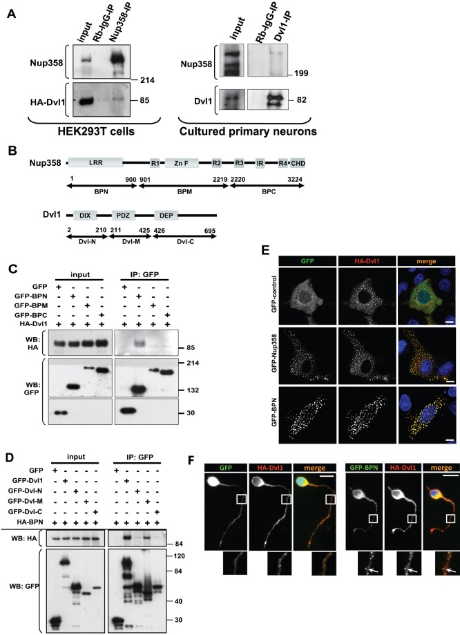
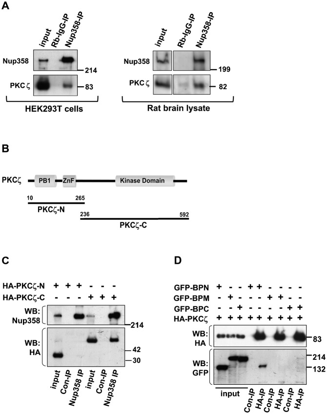
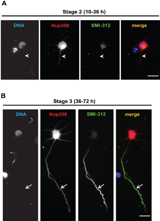
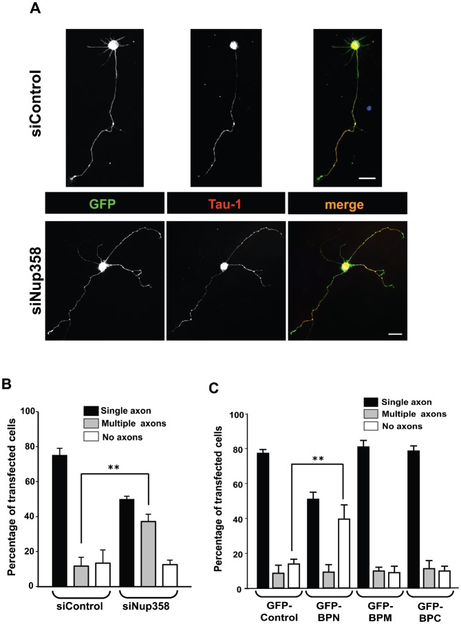
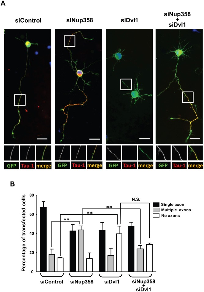
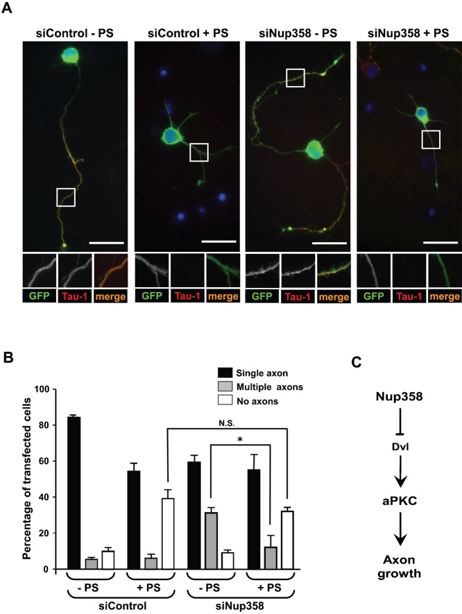
Par polarity complex, consisting of Par3, Par6, and aPKC, plays a conserved role in the establishment and maintenance of polarization in diverse cellular contexts. Recent reports suggest that Dishevelled (Dvl), a cytoplasmic mediator of Wnt signalling, interacts with atypical protein kinase C and regulates its activity during neuronal differentiation and directed cell migration. Here we show that Nup358 (also called RanBP2), a nucleoporin previously implicated in polarity during directed cell migration, interacts with Dishevelled and aPKC through its N-terminal region (BPN) and regulates axon-dendrite differentiation of cultured hippocampal neurons. Depletion of endogenous Nup358 leads to generation of multiple axons, whereas overexpression of BPN abrogates the process of axon formation. Moreover, siRNA-mediated knockdown of Dvl or inhibition of aPKC by a pseudosubstrate inhibitor significantly reverses the multiple axon phenotype produced by Nup358 depletion. Collectively, these data suggest that Nup358 plays an important role in regulating neuronal polarization upstream to Dvl and aPKC.

Keywords: Dishevelled; Neuron; Nucleoporin; Nup358; Polarity; aPKC.


Conflict of interest statement

Competing interests: The authors have no competing interests to declare.

Figures

Fig. 1.
Fig. 1.. Nup358 interacts with Dishevelled.
(A) Left panel: HA-tagged Dvl1 was overexpressed in HEK293T cells and endogenous Nup358 was immunoprecipitated (IP) using rabbit polyclonal antibodies against Nup358 (Nup358-IP). Rabbit IgG (Rb-IgG-IP) was used as control. Immunoprecipitates were analysed by western blotting (WB) with antibodies against HA and mAb414 (mouse monoclonal antibody that recognizes nucleoporins possessing FxFG sequences such as Nup358). Molecular weight markers are as indicated (in kDa). Right panel: Cultured primary neurons isolated from E18 rat were subjected to immunoprecipitation using Dvl1 antibody. The immunoprecipitate was probed with Dvl1 and Nup358 antibodies. (B) Domain structure of full-length Nup358 and Dvl1 and their fragments used in the study. The numbers refer to the amino acid positions. (C) HEK293T cells co-expressing HA-Dvl1 and GFP-control or GFP-tagged fragments of Nup358 were subjected to immunoprecipitation using anti-GFP antibodies. The immunoprecipitates were probed with antibodies against GFP or HA. (D) GFP-tagged full-length or fragments of Dvl1 were co-expressed with HA-BPN and were immunoprecipitated using anti-GFP antibodies. The immunoprecipitates were subjected to western analysis using indicated antibodies. (E) COS-7 cells were co-transfected with HA-Dvl1 (red) and GFP control, GFP-Nup358 or GFP-BPN (green) and were immunostained with anti-HA antibodies. DNA was visualized by Hoechst 33342 staining (blue). Scale bar, 10 µm. (F) E18 rat hippocampal neurons were transfected with pBetaActin-eGFP (GFP, green) or pBetaActin-BPN-eGFP (GFP-BPN, green) and pBetaActin-HA-Dvl1 (HA-Dvl1, red) constructs for 72 hours. The cells were fixed, stained and analyzed by fluorescence microscopy. DNA was stained with Hoechst 33342 dye. Scale bar, 20 µm. Arrows indicate co-localization of GFP-BPN with HA-Dvl1 puncta in neuronal extensions.
Fig. 2.
Fig. 2.. Nup358 interacts with aPKC.
(A) Left panel: HEK293T was subjected to immunoprecipitation using rabbit polyclonal antibodies against Nup358 (Nup358-IP) or control rabbit IgG (Rb-IgG-IP), and the immunoprecipitates were immunoblotted using indicated antibodies. Right panel: E18 rat brain lysate was subjected to immunoprecipitation using Nup358 antibody. The immunoprecipitate was probed with aPKC (PKCζ) and Nup358 antibodies. (B) The domain structure of PKCζ and the fragments used in the study. (C) Lysates of HEK293T cells expressing HA-tagged fragments of PKCζ were subjected to immunoprecipitation using control Rb-IgG (Rb-IgG-IP) or anti-Nup358 (Nup358-IP) antibodies. The immunoprecipitates were probed with HA-specific antibodies and MAb414 (for Nup358). (D) Cells were co-transfected with HA-PKCζ and GFP-tagged version of BPN, BPM or BPC, and the lysates were subjected to immunoprecipitation using EZview control (Con-IP) or EZview HA (HA-IP) beads (Sigma–Aldrich). Immunoblotting was performed using indicated antibodies.
Fig. 3.
Fig. 3.. Localization of endogenous Nup358 in polarizing rat hippocampal neurons.
Cultured rat hippocampal neurons, at stage 2 (A) and stage 3 (B), were immunostained with Nup358 (red) and SMI-312 (axonal marker; green). Arrow heads indicate immature neurites at stage 2 (B). Arrows indicate axons as identified SMI-312 axonal marker. DNA was visualized by Hoechst 33342 staining (blue). Scale bar, 25 µm.
Fig. 4.
Fig. 4.. Nup358 is required for neuronal polarization.
(A) E18 hippocampal neurons were transfected with control (siControl) or Nup358 siRNA (siNup358) along with pBetaActin-eGFP as transfection control, and were immunostained after culturing for 72 hours in vitro. The transfected neurons were identified by GFP expression (green). The effect of Nup358 depletion on axon formation was analysed using anti-Tau-1 antibodies (red). Arrows indicate axons as identified by Tau-1 axonal marker. DNA was stained with Hoechst 33342 dye (blue). Scale bar, 25 µm. (B) Quantitative analysis of the effect of Nup358 depletion on neuronal polarity. Error bars indicate standard deviations, n = 3, **P<0.01, Student's t test. (C) E18 rat hippocampal neurons were transfected with GFP-control, GFP tagged version of BPN, BPM or BPC and assessed for the effect on neuronal polarization after 72 hours. Error bars indicate standard deviations, n = 3, **P<0.01, Student's t test.
Fig. 5.
Fig. 5.. Nup358 functions upstream of Dvl.
(A) Hippocampal neurons were transfected with control siRNA (siControl), Nup358 siRNA (siNup358), Dvl1 siRNA (siDvl1) alone or co-transfected with siNup358 and siDvl1 as indicated. pBetaActin-eGFP (green) was used as the transfection marker. The transfected cells were stained with Tau-1 (red) to study the effect on axon formation. DNA was stained with Hoechst 33342 dye (blue). Scale bar, 25 µm. (B) Quantitative analysis of the effect of siRNA mediated depletion of different proteins on axon formation. Error bars indicate standard deviations, n = 3, **P<0.01, N.S. – non-significant, Student's t test.
Fig. 6.
Fig. 6.. Multiple axons formed by Nup358 depletion is partially rescued by inhibition of aPKC activity.
(A) Hippocampal neurons transfected with control (siControl) or Nup358 (siNup358) siRNA along with pBetaActin-eGFP and were cultured in the presence (+ PS) or absence (− PS) a pseudosubstrate inhibitor specific for PKCζ (10 µM). The neurons were fixed and immunostained after 72 hours. The effect of different treatments on neuronal polarization was assessed by scoring for GFP expression (green) and Tau-1 staining (red). Scale bar, 25 µm. (B) The quantitative analysis of the effect on neuronal polarity. Error bars indicate standard deviations, n = 3, *P<0.05, Student's t test. (C) A working model for the function of Nup358 in neuronal polarization. During the initial stages of single axon formation, Dvl-mediated activation of aPKC occurs at the nascent axon. Differentiation of other neurites into axons could be spatially and temporally inhibited by Nup358-mediated interference of Dvl and aPKC functions.

Similar articles

Cited by

References

    1. Arimura N., Kaibuchi K. (2007). Neuronal polarity: from extracellular signals to intracellular mechanisms. Nat. Rev. Neurosci. 8, 194–205 10.1038/nrn2056 - DOI - PubMed
    1. Barnes A. P., Polleux F. (2009). Establishment of axon–dendrite polarity in developing neurons. Annu. Rev. Neurosci. 32, 347–381 10.1146/annurev.neuro.31.060407.125536 - DOI - PMC - PubMed
    1. Conde C., Cáceres A. (2009). Microtubule assembly, organization and dynamics in axons and dendrites. Nat. Rev. Neurosci. 10, 319–332 10.1038/nrn2631 - DOI - PubMed
    1. D'Angelo M. A., Hetzer M. W. (2008). Structure, dynamics and function of nuclear pore complexes. Trends Cell Biol. 18, 456–466 10.1016/j.tcb.2008.07.009 - DOI - PMC - PubMed
    1. Etemad-Moghadam B., Guo S., Kemphues K. J. (1995). Asymmetrically distributed PAR-3 protein contributes to cell polarity and spindle alignment in early C. elegans embryos. Cell 83, 743–752 10.1016/0092-8674(95)90187-6 - DOI - PubMed