

Right salpingo-ovarian and distal ileal entrapment within a paracaecal hernia presenting as acute appendicits
- PMID: 24246293
- PMCID: PMC3860031
- DOI: 10.1016/j.ijscr.2013.10.007
Right salpingo-ovarian and distal ileal entrapment within a paracaecal hernia presenting as acute appendicits
Abstract
Introduction: Pericaecal hernias are a rare subgroup of internal abdominal hernias that present with abdominal pain and occasionally with features of bowel obstruction.
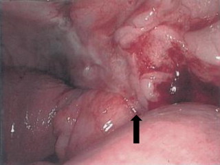
Presentation of case: A 72 year old female presented with a 24-h history of sharp, localised right iliac fossa pain, and no other symptoms. Clinical examination confirmed localised peritonism in the right iliac fossa. A tentative diagnosis of acute appendicitis was considered but in view of age a CT scan was performed. An area of abnormality in the right iliac fossa region was noted. At laparoscopy a macroscopically normal appendix and caecum was found. A smooth non-indentable mass in the lateral right iliac fossa contained loops of distal ileum, passing through a retro-caecal mesenteric defect consistent with a paraceacal hernia, with entrapment of the right ovary and fallopian tube. A right salpingectomy as performed and subsequent histopathological examination confirmed infarction of the fallopian tube.
Discussion: Internal abdominal hernias are reported to have a post mortem incidence ranging between 0.2 and 0.9% of which only 10-15% are accounted for by pericaecal hernias. Types of pericaecal hernias include: ileocolic, retrocaecal, ileocaecal and paracaecal. These hernias are predisposed by the embryological development of the caecum retracting to the posterior abdominal wall and forming potential fossae.
Conclusion: This case highlights the need to consider a pericaecal hernia as a differential cause of right iliac fossa peritonism, and an indication for radiological imaging such as CT scan when the history is atypical for acute appendicitis.
Keywords: Appendicitis; Ileum entrapment; Paracaecal hernia; Salpingo-ovarian.
Copyright © 2013 The Authors. Published by Elsevier Ltd.. All rights reserved.
Figures
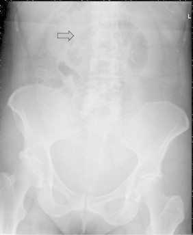
 ).
).
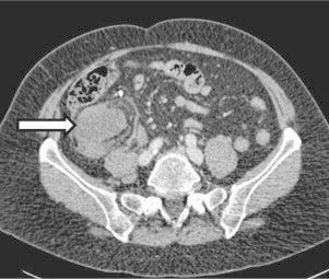
 ) with the caecum displaced anteriorly.
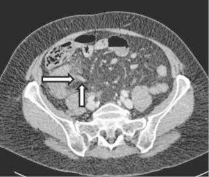 ) in the peritoneal recess and displacement of the mesenteric vascular pedicle (
 ). Unable to positively identify the appendix.
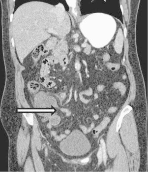 ) in the peritoneal recess
References
-
- Ghahremani G.G. Internal abdominal hernia. Surgical Clinics of North America. 1984;643:93–406. - PubMed
-
- Choh N.A., Rasheed M., Jehangir M. The computed tomography diagnosis of paracecal hernia. Hernia. 2010;14(5):527–529. - PubMed
-
- Larson W.J. 3rd ed. Churchill Livingstone; Philadelphia, PA: 2001. Human embryology.
-
- Meyers M. Internal abdominal hernias. In: Meyers M.A., editor. Dynamic radiology of the abdomen: normal and pathologic anatomy. 5th ed. Springer; New York: 2000. pp. 711–748.
-
- Mathieu D., Luciano A. Internal abdominal herniations. American Journal of Roentology. 2004;183:397–404. - PubMed
LinkOut - more resources
Full Text Sources
Other Literature Sources
Research Materials
