. 2014 Jan 22;179(100):93-101.
doi: 10.1016/j.virusres.2013.11.006. Epub 2013 Nov 15.

Changes in immune cell populations in the periphery and liver of GBV-B-infected and convalescent tamarins (Saguinus labiatus)

Affiliations

Changes in immune cell populations in the periphery and liver of GBV-B-infected and convalescent tamarins (Saguinus labiatus)

Simon P Hood et al. Virus Res. .

Abstract

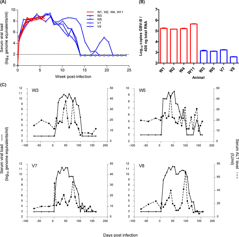
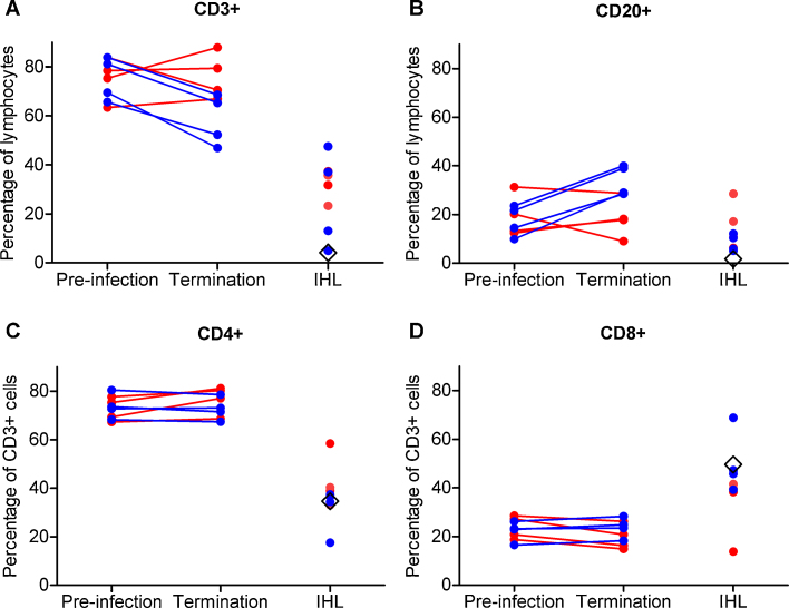
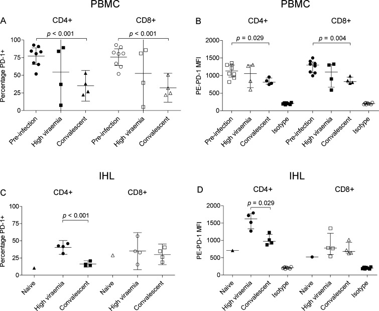
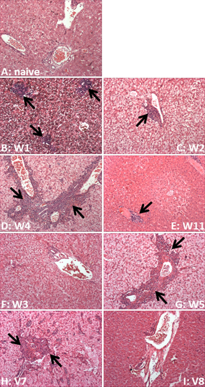
Flaviviruses related to hepatitis C virus (HCV) in suitable animal models may provide further insight into the role that cellular immunity contributes to spontaneous clearance of HCV. We characterised changes in lymphocyte populations in tamarins with an acute GBV-B infection, a hepatitis virus of the flaviviridae. Major immune cell populations were monitored in peripheral and intra-hepatic lymphocytes at high viraemia or following a period when peripheral virus was no longer detected. Limited changes in major lymphocyte populations were apparent during high viraemia; however, the proportions of CD3(+) lymphocytes decreased and CD20(+) lymphocytes increased once peripheral viraemia became undetectable. Intrahepatic lymphocyte populations increased at both time points post-infection. Distinct expression patterns of PD-1, a marker of T-cell activation, were observed on peripheral and hepatic lymphocytes; notably there was elevated PD-1 expression on hepatic CD4(+) T-cells during high viraemia, suggesting an activated phenotype, which decreased following clearance of peripheral viraemia. At times when peripheral vRNA was not detected, suggesting viral clearance, we were able to readily detect GBV-B RNA in the liver, indicative of long-term virus replication. This study is the first description of changes in lymphocyte populations during GBV-B infection of tamarins and provides a foundation for more detailed investigations of the responses that contribute to the control of GBV-B infection.

Keywords: 4-(2-hydroxyethyl)-1-piperazineethanesulfonic acid; APC; Acute viral hepatitis; CTLA4; DMSO; EGTA; FITC; Fluorescein isothiocyanate; GB virus B; GBV-B; HBSS; HCV; HEPES; Hank's balanced salt solution; IFN; IHL; IVT; Immune cell; MFI; MHC; NK; NS; PD-1; PD1-L1; PE; RPMI; Roswell Park Memorial Institute medium; allophycocyanin; cytotoxic T lymphocyte antigen-4; dimethyl sulphoxide; ethylene glycol tetraacetic acid; ge; genome equivalents; hepatitis C virus; in vitro transcription; interferon; intrahepatic lymphocytes; major histocompatibility complex; median fluorescence intensity; natural killer; non-structural; phycoerythrin; programmed death receptor-1; programmed death receptor-1 ligand; qRT-PCR; quantitative reverse transcriptase polymerase chain reaction; vRNA; viral ribonucleic acid.


Figures

Supplementary Fig. S1
Expression of PD-1 on peripheral (top panels) and intrahepatic (bottom panels) CD3+CD4+ lymphocytes. Representative histograms from animals terminated during acute viremia (W4, (A) PBMC, (C) IHL), convalescent phase (W3, (B) PBMC, (D) IHL) and naive animal (G20, (E) IHL). Brown, isotype control antibody; black, pre-infection PBMC; red, viremic phase samples; blue, convalescent phase samples. (F) Gating strategy for analysis of PD-1 positive lymphocytes showing a representative sample stained with isotype control (bottom centre) or PD-1 specific antibody (bottom right).
Supplementary Fig. S2
Cellular immune responses against GBV-B NS3 in IHL isolated at time of termination and stimulated with the indicated peptide pools or PMA as positive control. Bars indicate percentage of CD4+ (left panel) or CD8+ (right panel) T-cells expressing IFNg following stimulation. All values are background (DMSO stimulation) subtracted. Dashed line indicates assay cutoff, defined as the mean of all negative samples plus three standard deviations. Red text indicates animals terminated at a time of high viremia (6 wpi); blue text indicates animals terminated following clearance of detectable GBV-B infection.
Fig. 1
Fig. 1
Dynamics of GBV-B infection in red-bellied tamarins. (A) Viral load was monitored at weekly intervals in eight animals inoculated with 107 genome equivalents of infectious serum. Red lines represent animals terminated at 6 weeks post-infection. Animals displayed uniform viraemia profiles; hence symbols are omitted for clarity; blue lines indicate animals terminated in the convalescent phase. Dashed line indicates the limit of quantification of the qRT-PCR assay (100 ge/ml serum). (B) Viral load in the liver is presented as copies of virus per 400 ng total RNA (approximately 10,000–15,000 cells) for each animal. The limit of quantification was 7.6 × 10−2/400 ng total RNA. Red bars indicate the animals terminated at 6 wpi; blue bars indicate the animals terminated in convalescence. (C) Changes in serum liver enzyme levels during infection are shown for the four animals which proceeded to convalescence. ALT levels (dotted line) are indicated for pre- and post-infection time points alongside peripheral viral load (solid line).
Fig. 2
Fig. 2
Peripheral and hepatic lymphocyte dynamics during GBV-B infection of red-bellied tamarins. Percentage of lymphocyte fraction (as determined by forward scatter and side scatter characteristics) positive for (A) CD3 and (B) CD20. Percentage of CD3+ fraction positive for (C) CD4 and (D) CD8. Animals are grouped according to the phase of infection at which they were sacrificed. Lymphocytes from a naive animal, G20, are represented by a black diamond.
Fig. 3
Fig. 3
Analysis of PD-1 expression on peripheral and intrahepatic CD3+ lymphocytes from GBV-B-infected tamarins. (A and C) Proportion of CD4+ and CD8+ cells positive for PD-1 expression in PBMC and IHL prior to infection, during the viremic (6 wpi) and convalescent phase (24 wpi). (B and D) Median fluorescence intensity (MFI) of PE-PD-1 stained CD4+ and CD8+ populations. Naïve, cells from naive animal G20; Isotype, MFI of cells stained with PE-labelled isotype-matched control antibody; error bars indicate mean and 95% confidence intervals (left panels) or median and interquartile range (right panels). Significant differences between groups are indicated by relevant p values.
Fig. 4
Fig. 4
Haematoxylin and eosin staining of liver tissue. (A) Naive tamarin; (B–E) tamarins terminated at high viraemia; (F–I) tamarins terminated at convalescence. Lymphoctye infiltration is indicated by arrows. Magnification 100×.

Similar articles

Cited by

References

    1. Barber D.L., Wherry E.J., Masopust D., Zhu B., Allison J.P., Sharpe A.H., Freeman G.J., Ahmed R. Restoring function in exhausted CD8T cells during chronic viral infection. Nature. 2006;439:682–687. - PubMed
    1. Beames B., Chavez D., Guerra B., Notvall L., Brasky K.M., Lanford R.E. Development of a primary tamarin hepatocyte culture system for GB virus-B: a surrogate model for hepatitis C virus. J. Virol. 2000;74:11764–11772. - PMC - PubMed
    1. Beames B., Chavez D., Lanford R.E. GB virus B as a model for hepatitis C virus. ILAR J. 2001;42:152–160. - PubMed
    1. Bowen D.G., Walker C.M. Adaptive immune responses in acute and chronic hepatitis C virus infection. Nature. 2005;436:946–952. - PubMed
    1. Bright H., Carroll A.R., Watts P.A., Fenton R.J. Development of a GB virus B marmoset model and its validation with a novel series of hepatitis C virus NS3 protease inhibitors. J. Virol. 2004;78:2062–2071. - PMC - PubMed

Publication types