

The α1 adrenoceptor antagonist prazosin enhances sleep continuity in fear-conditioned Wistar-Kyoto rats
- PMID: 24246572
- PMCID: PMC3969852
- DOI: 10.1016/j.pnpbp.2013.11.004
The α1 adrenoceptor antagonist prazosin enhances sleep continuity in fear-conditioned Wistar-Kyoto rats
Abstract
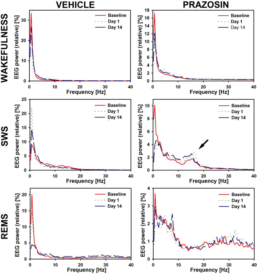
Fragmentation of rapid eye movement sleep (REMS) is well described in individuals with posttraumatic stress disorder (PTSD) and likely has significant functional consequences. Fear-conditioned rodents may offer an attractive model of the changes in sleep that characterize PTSD. Following fear conditioning (FC), Wistar-Kyoto (WKY) rats, a strain known to be particularly stress-sensitive, have increased REMS fragmentation that can be quantified as a shift in the distribution of REMS episodes towards the more frequent occurrence of sequential REMS (inter-REMS episode interval≤3 min) vs. single REMS (interval>3 min). The α1 adrenoceptor antagonist prazosin has demonstrated efficacy in normalizing sleep in PTSD. To determine the utility of fear-conditioned WKY rats as a model of sleep disturbances typical of PTSD and as a platform for the development of new treatments, we tested the hypothesis that prazosin would reduce REMS fragmentation in fear-conditioned WKY rats. Sleep parameters and freezing (a standard measure of anxiety in rodents) were quantified at baseline and on Days 1, 7, and 14 following FC, with either prazosin (0.01mg/kg, i.p.) or vehicle injections administered prior to testing in a between-group design. Fear conditioning was achieved by pairing tones with a mild electric foot shock (1.0mA, 0.5s). One, 7, and 14 days following FC, prazosin or vehicle was injected, the tone was presented, freezing was measured, and then sleep was recorded from 11 AM to 3 PM. WKY rats given prazosin, compared to those given vehicle, had a lower amount of seq-REMS relative to total REMS time 14 days after FC. They also had a shorter non-REMS latency and fewer non-REMS arousals at baseline and on Days 1 and 7 after FC. Thus, in FC rats, prazosin reduced both REMS fragmentation and non-REMS discontinuity.
Keywords: BLA; BNST; CNA; CS; ECG; EEG; EMG; FC; Fear conditioning; LA; NE; Norepinephrine; PTSD; Posttraumatic stress disorder; REM sleep; REMS; VLPO; WKY; Wistar–Kyoto; Wistar–Kyoto rats; basolateral nucleus of the amygdala; bed nucleus of the stria terminalis; central nucleus of the amygdala; conditioning stimulus; electrocardiogram; electroencephalogram; electromyogram; fear-conditioned or fear conditioning; lateral nucleus of the amygdala; norepinephrine; posttraumatic stress disorder; rapid eye movement sleep; seq-REMS; sequential rapid eye movement sleep; si-REMS; single rapid eye movement sleep; ventrolateral preoptic nucleus.
Published by Elsevier Inc.
Figures


References
-
- Amaral D, Price J, Pitkanen A, Carmichael S. Anatomical organization of the primate amygdaloid complex. In: Aggleton J, editor. The Amygdala: Neurobiological Aspects of Emotion, Memory, and Mental Dysfunction. New York: Wiley-Liss, Inc.; 1992.
-
- American Sleep Disorders Association. EEG arousals: scoring rules and examples: a preliminary report from the Sleep Disorders Atlas Task Force of the American Sleep Disorders Association. Sleep. 1992;15:173–184. - PubMed
-
- Amici R, Zamboni G, Perez E, Jones CA, Toni I, Culin F, et al. Pattern of desynchronized sleep during deprivation and recovery induced in the rat by changes in ambient temperature. J Sleep Res. 1994;3:250–256. - PubMed
-
- Becker ME, Hertzberg MA, Moore SD, Dennis MF, Bukenya DS, Beckham JC. A placebo-controlled trial of bupropion SR in the treatment of chronic posttraumatic stress disorder. J Clin Psychopharmacol. 2007;27:193–197. - PubMed
Publication types
MeSH terms
Substances
Grants and funding
LinkOut - more resources
Full Text Sources
Other Literature Sources
