Review
. 2013 Oct;50(4):348-59.
doi: 10.1053/j.seminhematol.2013.09.001.

Detectable clonal mosaicism in the human genome

Affiliations
Review

Detectable clonal mosaicism in the human genome

Mitchell J Machiela et al. Semin Hematol. 2013 Oct.

Abstract

Human genetic mosaicism is the presence of two or more cellular populations with distinct genotypes in an individual who developed from a single fertilized ovum. While initially observed across a spectrum of rare genetic disorders, detailed assessment of data from genome-wide association studies now reveal that detectable clonal mosaicism involving large structural alterations (>2 Mb) can also be seen in populations of apparently healthy individuals. The first generation of descriptive studies has generated new interest in understanding the molecular basis of the affected genomic regions, percent of the cellular subpopulation involved, and developmental timing of the underlying mutational event, which could reveal new insights into the initiation, clonal expansion, and phenotypic manifestations of mosaic events. Early evidence indicates detectable clonal mosaicism increases in frequency with age and could preferentially occur in males. The observed pattern of recurrent events affecting specific chromosomal regions indicates some regions are more susceptible to these events, which could reflect inter-individual differences in genomic stability. Moreover, it is also plausible that the presence of large structural events could be associated with cancer risk. The characterization of detectable genetic mosaicism reveals that there could be important dynamic changes in the human genome associated with the aging process, which could be associated with risk for common disorders, such as cancer, cardiovascular disease, diabetes, and neurological disorders.


Figures

Figure 1
Figure 1
Two example homologous chromosomes are depicted in the nucleus of a diploid individual for a cellular population. The underlying process of mosaicism is depicted where an individual with a normal karyotype carrying a complete paternal (white) and maternal (black) chromosome in all cells undergoes a mutational event that clonally expands to become a detectible subset of the total cellular population. This can result in a mosaic loss on the maternal chromosome in only a portion of the cellular population (orange), a mosaic gain on the maternal chromosome (yellow), or a mosaic copy neutral loss of heterozygosity (black) such as an acquired uniparental disomy.
Figure 2
Figure 2
Illustrations of technologies used to detect mosaicism. (A) Karyotype as visualized using light microscopy and staining to visualize banding patterns. (B) “Painted” karyotype using SKY tagged probes. (C) Sanger sequencing reads from radioisotope labeled gel (left) and fluorescent labeled chromatogram (right). (D) Array CGH with comparative copy loss and gain on three example chromosomes. (E) BAF and LRR values extracted from SNP array plotting an example copy gain on a chromosome. (F) Next-generation sequencing reads generated for a chromosomal region of interest.
Figure 2
Figure 2
Illustrations of technologies used to detect mosaicism. (A) Karyotype as visualized using light microscopy and staining to visualize banding patterns. (B) “Painted” karyotype using SKY tagged probes. (C) Sanger sequencing reads from radioisotope labeled gel (left) and fluorescent labeled chromatogram (right). (D) Array CGH with comparative copy loss and gain on three example chromosomes. (E) BAF and LRR values extracted from SNP array plotting an example copy gain on a chromosome. (F) Next-generation sequencing reads generated for a chromosomal region of interest.
Figure 3
Figure 3
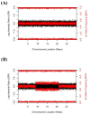
Simulated plots of GWAS SNP array data depicting (A) a normal chromosome, (B) a chromosome exhibiting an acquired mosaic uniparental disomy, (C) chromosome with a mosaic gain, and (D) a chromosome with a mosaic loss. Log 2 R ratios (LRR) are points plotted in black on the left y axis and give an indication of copy number (gain>0, loss<0). B allele frequencies (BAF) are points plotted in red on the right y axis and are indicators of allelic imbalance. Each SNP on the genotyping array has a LRR (black) and BAF (red) point plotted at the SNP’s chromosomal position along the x axis.
Figure 3
Figure 3
Simulated plots of GWAS SNP array data depicting (A) a normal chromosome, (B) a chromosome exhibiting an acquired mosaic uniparental disomy, (C) chromosome with a mosaic gain, and (D) a chromosome with a mosaic loss. Log 2 R ratios (LRR) are points plotted in black on the left y axis and give an indication of copy number (gain>0, loss<0). B allele frequencies (BAF) are points plotted in red on the right y axis and are indicators of allelic imbalance. Each SNP on the genotyping array has a LRR (black) and BAF (red) point plotted at the SNP’s chromosomal position along the x axis.

References

    1. Zlotnikoff M. A Human Mosaic. J Heredity. 1945;36:163–8.
    1. Strachan T, Read AP, Strachan T. Human molecular genetics. 4. New York: Garland Science; 2011. p. xxv.p. 781.
    1. Larsson NG. Somatic mitochondrial DNA mutations in mammalian aging. Annu Rev Biochem. 2010;79:683–706. - PubMed
    1. Bianchi NO, Bianchi MS, Richard SM. Mitochondrial genome instability in human cancers. Mutat Res. 2001;488:9–23. - PubMed
    1. Taylor RW, Turnbull DM. Mitochondrial DNA mutations in human disease. Nat Rev Genet. 2005;6:389–402. - PMC - PubMed