Review
. 2014 Apr;35(2):197-220.
doi: 10.1016/j.yfrne.2013.11.001. Epub 2013 Nov 16.

Gonadal steroid hormones and the hypothalamo-pituitary-adrenal axis

Affiliations
Review

Gonadal steroid hormones and the hypothalamo-pituitary-adrenal axis

Robert J Handa et al. Front Neuroendocrinol. 2014 Apr.

Abstract

The hypothalamo-pituitary-adrenal (HPA) axis represents a complex neuroendocrine feedback loop controlling the secretion of adrenal glucocorticoid hormones. Central to its function is the paraventricular nucleus of the hypothalamus (PVN) where neurons expressing corticotropin releasing factor reside. These HPA motor neurons are a primary site of integration leading to graded endocrine responses to physical and psychological stressors. An important regulatory factor that must be considered, prior to generating an appropriate response is the animal's reproductive status. Thus, PVN neurons express androgen and estrogen receptors and receive input from sites that also express these receptors. Consequently, changes in reproduction and gonadal steroid levels modulate the stress response and this underlies sex differences in HPA axis function. This review examines the make up of the HPA axis and hypothalamo-pituitary-gonadal (HPG) axis and the interactions between the two that should be considered when exploring normal and pathological responses to environmental stressors.

Keywords: Androgen receptor; CRF; Dihydrotestosterone; Estradiol; Estrogen receptor; Glucocorticoid; HPA; HPG; Sex difference; Testosterone.


Figures

Figure 1
Figure 1
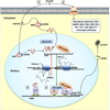
Diagrammatic representation of the various Intracellular actions of steroid hormone receptors. (1) Steroids can freely diffuse through the plasma membrane lipid bilayer and bind to a steroid hormone receptor (SHR) associated with heat shock protein (HSP90) in the cytoplasm or nucleus. Steroid binding leads to the release of HSP90 and translocation of the SHR to chromatin DNA. (2) SHR homo- or heterodimers recruit coregulatory proteins such as the p160 and p300 family of coactivators. These coactivators have intrinsic histone acetyl transferase (HAT) activity and can acetylate histones associated with inactive chromatin to uncoil DNA and expose regulatory regions and transcription initiation sites of target genes. The SHR dimer then binds to a regulatory region (e.g., hormone response element, HRE) and in association with these coregulatory protein factors (e.g., SRC, CBP) can stimulate transcription through activation of the pre-initiation complex (PIC). Transcription can subsequently be inhibited by the activity of histone deacetylases (HDACs), which act to remove acetyl groups from histones and promote chromatin recoiling. (3) Steroid-bound receptor can also act as a coactivator or corepressor and affect gene expression by tethering to other DNA bound transcription factors (e.g. fos and jun at an AP-1 response element). (4) Steroids have effects attributable to actions at a membrane-associated steroid hormone receptor (mSHR). These receptors can be classic SHRs tethered to the membrane, G-protein coupled receptors, or ion channels. These effects are diverse and include alterations in membrane potential and second messenger pathways. AP-1 RE, activator protein 1 response element; Ca2+, calcium; CBP, cyclic adenosine monophosphate response element binding protein (CREB) binding protein; ERK, extracellular regulated kinase; HAT, histone acetyl transferase; HDAC, histone deacetylase; HRE, hormone response element; HSP90, heat shock protein 90; MAPK, mitogen-activated protein kinase; mSHR, membrane-associated steroid hormone receptor; NOS, nitric oxide synthase; PIC, pre-initiation complex; PKA, protein kinase A; PKB, protein kinase B; PKC, protein kinase C; PLC, phospholipase C; SHR, steroid hormone receptor; SRC, steroid receptor coactivator; TATA, core promoter sequence.
Figure 2
Figure 2
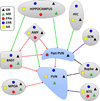
Distribution of gonadal and adrenal steroid hormone receptors in relationship to inputs to HPA circuitry. Gonadal and adrenal steroid hormone receptors have considerable overlap in expression within brain regions that have direct inputs to the paraventricular nucleus (PVN) and peri-PVN. The brain regions involved and the overall influence on the output of the PVN depends upon stressor modality (psychogenic, limbic; homeostatic, brain stem) and gonadal steroid hormone levels derived from systemic and/or local de novo sources. Green solid arrows indicate excitatory glutamate connections. Red dashed arrows indicate inhibitory gamma-aminobutyric acid (GABA) connections. Black arrow indicates serotonergic connections. Blue arrow indicates mixed norepinephrine and glucagon-like peptide 1 connections. Purple arrow indicates mixed GABA and arginine vasopressin connections. AMY, amygdala; AR, androgen receptor; BNST, bed nucleus of the stria terminalis; ERα, estrogen receptor alpha; ERβ, estrogen receptor beta; GR, glucocorticoid receptor; MPOA, medial preoptic area; MR, mineralocorticoid receptor; NTS, nucleus of the solitary tract; PFC, prefrontal cortex; PVN, paraventricular nucleus; Raphe, dorsal raphe nucleus; SCN, suprachiasmatic nucleus.

References

    1. Abel K, Majzoub JA. Molecular biology of the HPA axis. In: Steckler T, Kalin NH, Reul JM, editors. Handbook of Stress and the Brain. Vol. 15. Elsevier; 2005. pp. 79–94.
    1. Abraham IM, Todman MG, Korach KS, Herbison AE. Critical in vivo roles for classical estrogen receptors in rapid estrogen actions on intracellular signaling in mouse brain. Endocrinology. 2004;145:3055–3061. - PubMed
    1. Adan RA, Cox JJ, Beischlag TV, Burbach JP. A composite hormone response element mediates the transactivation of the rat oxytocin gene by different classes of nuclear hormone receptors. Mol Endocrinol. 1993;7:47–57. - PubMed
    1. Adler A, Vescovo P, Robinson JK, Kritzer MF. Gonadectomy in adult life increases tyrosine hydroxylase immunoreactivity in the prefrontal cortex and decreases open field activity in male rats. Neuroscience. 1999;89:939–954. - PubMed
    1. Aguilera G, Nikodemova M, Wynn PC, Catt KJ. Corticotropin releasing hormone receptors: two decades later. Peptides. 2004;25:319–329. - PubMed

MeSH terms

Substances

LinkOut - more resources