. 2014 Jan 3;289(1):152-62.
doi: 10.1074/jbc.M113.459271. Epub 2013 Nov 18.

Pseudomonas aeruginosa Cif protein enhances the ubiquitination and proteasomal degradation of the transporter associated with antigen processing (TAP) and reduces major histocompatibility complex (MHC) class I antigen presentation

Affiliations

Pseudomonas aeruginosa Cif protein enhances the ubiquitination and proteasomal degradation of the transporter associated with antigen processing (TAP) and reduces major histocompatibility complex (MHC) class I antigen presentation

Jennifer M Bomberger et al. J Biol Chem. .

Abstract

Cif (PA2934), a bacterial virulence factor secreted in outer membrane vesicles by Pseudomonas aeruginosa, increases the ubiquitination and lysosomal degradation of some, but not all, plasma membrane ATP-binding cassette transporters (ABC), including the cystic fibrosis transmembrane conductance regulator and P-glycoprotein. The goal of this study was to determine whether Cif enhances the ubiquitination and degradation of the transporter associated with antigen processing (TAP1 and TAP2), members of the ABC transporter family that play an essential role in antigen presentation and intracellular pathogen clearance. Cif selectively increased the amount of ubiquitinated TAP1 and increased its degradation in the proteasome of human airway epithelial cells. This effect of Cif was mediated by reducing USP10 deubiquitinating activity, resulting in increased polyubiquitination and proteasomal degradation of TAP1. The reduction in TAP1 abundance decreased peptide antigen translocation into the endoplasmic reticulum, an effect that resulted in reduced antigen available to MHC class I molecules for presentation at the plasma membrane of airway epithelial cells and recognition by CD8(+) T cells. Cif is the first bacterial factor identified that inhibits TAP function and MHC class I antigen presentation.

Keywords: ABC Transporter; Airway Epithelial Cell; Antigen Processing; Deubiquitination; Pseudomonas aeruginosa; TAP; Ubiquitination; Viral-Bacterial Co-infection.


Figures
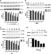
FIGURE 1.
FIGURE 1.
P. aeruginosa Cif induces the degradation of TAP1. a, OMVs isolated from Cif-expressing P. aeruginosa (subsequently called (+Cif) OMVs) applied apically to polarized airway epithelial cells induce a time-dependent reduction in TAP1, but not TAP2, protein abundance, as assessed by Western blot analysis. Ezrin is a protein-loading control, and TAP data for each lane are normalized for ezrin abundance and presented as a percentage of control (0 min, no OMV treatment). Similar results were obtained with Cif in A549 cells (data not shown). b, (−Cif) OMVs from P. aeruginosa do not reduce TAP1 protein levels. (+Cif) OMVs and (−Cif) OMVs were applied to the apical side of airway epithelial cells, and TAP1 protein abundance was assessed by Western blot analysis. Both representative blots shown were probed for TAP1. Quantification for Western blot experiments is presented below representative blots. c, recombinant Cif protein reduces TAP1 protein levels. Recombinant Cif protein (50 μg) was applied apically to airway epithelial cells for various times and TAP protein abundance assessed by Western blot analysis. Ezrin was used as a loading control. Quantification for Western blot experiments is found below representative blots. d, Cif reduces TAP1 abundance in a dose-dependent manner. Recombinant Cif protein was applied to the apical side of airway epithelial cells at various doses for 90 min, and TAP1 protein abundance was assessed by Western blot analysis. Quantification for Western blot experiments is presented below representative blots. Data are expressed as the mean ± S.E. (error bars), n = 3. *, p < 0.05.
FIGURE 2.
FIGURE 2.
Cif reduces peptide translocation into the ER and MHC class I antigen presentation. a, translocation of 125I-labeled B27#3 glycopeptide-1 was measured in ER microsomes isolated from airway epithelial cells treated with (+Cif) OMVs or (−Cif) OMVs for 90 min. Data are expressed as the mean ± S.E. (error bars), n = 3. *, p < 0.05. b, (+Cif) OMVs significantly reduced viral antigen presentation, as assessed by flow cytometry. EL-4 cells were infected with a PR8/SIINFEKL influenza strain, which is endogenously processed and presented as Kb-SIINFEKL. Kb-SIINFEKL was detected at the cell surface with the 25.D1–16 antibody (specifically recognizing Kb-SIINFEKL complexes at the cell surface) and flow cytometry. Absolute mean fluorescence intensity (MFI) values were: (−Cif) OMVs, 19.00 ± 0.57; (+Cif) OMVs, 3.17 ± 0.29. c, (+Cif) OMVs, but not (−Cif) OMVs, reduced cell surface expression of MHC class I (Kb), as assessed by flow cytometry in EL-4 cells using an APC-conjugated H-2Kb antibody. Infection of EL-4 cells with PR8/SIINFEKL influenza did not alter cell surface MHC class I levels. (+Cif) OMVs and (+Cif) OMVs in the presence of influenza virus reduced cell surface MHC class I. (−Cif) OMVs had no effect on cell surface MHC class I. Absolute MFI values were: control, 781.33 ± 2.03; (+Cif) OMVs, 129.33 ± 1.45; (+Cif) OMVs + flu, 131.67 ± 54.33; (−Cif) OMVs, 1008.33 ± 9.02. d, (+Cif) OMVs dramatically reduced CD8+ T cell activation during influenza A/Japan/57 infection. MLE-Kd cells were infected with A/Japan/57 or PR8 influenza in the presence of (+Cif) OMVs or (−Cif) OMVs and combined with CD8+ T cells specific for the HA204–212 minimal epitope. CD8+ T cell activation (i.e. TNF-α release) was quantified by ELISA. A/PR/8/34 is a control influenza strain, which does not express the HA204–212 epitope. Data are expressed as the mean ± S.E., and all experiments were repeated at least three times. *, p < 0.05.
FIGURE 3.
FIGURE 3.
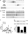
Cif trafficking to the ER via the retrograde pathway is required to reduce TAP1 abundance. Airway epithelial cells were lysed, and intracellular organelles were prepared via differential centrifugation and OptiPrep gradient separation. a, isolation of intracellular organelles. Plasma membrane (Na+,K+-ATPase), Golgi apparatus (TGN58), and ER (calnexin) resident proteins were used to identify by Western blot analysis the plasma membrane, Golgi apparatus, and ER, respectively. Representative blots of the isolated fractions are presented. b, OptiPrep gradient fractionation and differential centrifugation analysis of Cif (delivered via (+Cif) OMVs) at various times after incubation with airway epithelial cells reveals the sequential trafficking of Cif from the plasma membrane, to the Golgi, and then to the ER. Fractions correspond to those in a. Western blot analysis was performed for Cif. c, Cif protein detected in each fraction, as presented as representative blots in b, is quantified. Data are presented as percentage of Cif detected in each fraction compared with the total Cif detected in all fractions. d, inhibition of retrograde transport to the ER with brefeldin A (BrefA; 100 μm) inhibits the Cif-mediated decrease in TAP1 protein abundance, as assessed by Western blot analysis. n = 3; *, p < 0.05.
FIGURE 4.
FIGURE 4.
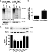
Cif increases the polyubiquitination status of TAP1 and promotes its proteasomal degradation. a, (+Cif) OMVs applied to airway epithelial cells elicit an increase in polyubiquitinated TAP1, as assessed by immunoprecipitation (IP) of TAP1 and Western blotting (IB) for polyubiquitin adducts with the FK1 ubiquitin antibody. Control cells were exposed to (−Cif) OMVs. IgG, immunoprecipitation using a nonimmune IgG was used as a specificity control. Black boxes highlight ubiquitinated TAP1 (please note that this experiment detects endogenous, ubiquitinated TAP, thus the signal is low compared with published studies that typically examine the ubiquitination of overexpressed proteins). 5% of the lysate was loaded in each lane labeled lysate, the remainder of the lysate was used for immunoprecipitation studies. b, quantification for Western blotting experiments is shown. Ubiquitinated TAP1 is normalized to the amount of TAP1 immunoprecipitated and is expressed as a percentage of the control (−Cif) OMV sample. c, proteasomal inhibitors (37.5 μm MG132) blocked the Cif-mediated degradation of TAP1, as assessed by Western blot analysis, whereas lysosomal inhibitors (200 μm chloroquine and 50 mm NH4Cl) had no effect. Experiments were performed in the presence of 20 μg/ml cycloheximide to prevent new protein synthesis. In each experiment samples treated with (+Cif) OMVs and (−Cif) OMVs were run on the same gel, but the gel images were cut for presentation. Quantification for Western blot experiments is presented with representative blots. Data are expressed as the mean ± S.E. (error bars), n = 3. *, p < 0.05.
FIGURE 5.
FIGURE 5.
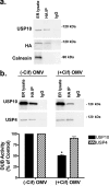
Cif inhibits USP10 activity associated with the endoplasmic reticulum. a, USP10 exhibits deubiquitinating activity in the ER fraction. Identification of DUBs covalently linked to the HA-UbVME probe was achieved by immunoprecipitation of the HA-UbVME-DUB complex using an anti-HA monoclonal antibody, followed by Western blot analysis. IgG, immunoprecipitation using a nonimmune IgG was used as a specificity control. Calnexin was present in the ER fraction but did not have DUB activity, as expected. Lysate lane represents 5% of total cell lysate. The remainder of the lysate was used for immunoprecipitation experiments. b, (+Cif) OMVs reduced the activity of a 110-kDa DUB in the ER as assessed by a DUB activity assay (see “Experimental Procedures,” Fig. 3A, and Refs. –17). ER lysate lane demonstrates USP10 protein abundance in ER fraction, whereas HA IP lane identifies active USP10 in the ER fraction. IgG, immunoprecipitation using a nonimmune IgG was used as a control. Lysate lane represents 5% of total cell lysate. The remainder of the lysate was used for immunoprecipitation experiments. In each experiment samples treated with (+Cif) OMVs and (−Cif) OMVs were run on the same gel, but the gel images were cut for presentation. Quantification for all Western blot experiments is presented. Data are expressed as the mean ± S.E. (error bars), n = 3. *. p < 0.05.
FIGURE 6.
FIGURE 6.
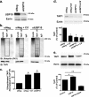
USP10 regulates the amount of ubiquitinated TAP1 and TAP1 abundance. a, siRNA (15 nm) knockdown of USP10 in airway epithelial cells, as assessed by Western blot analysis for USP10 and normalized for ezrin is shown. Representative blots are presented. b, siRNA knockdown of USP10 increased the amount of ubiquitinated TAP1, as assessed by immunoprecipitation (IP) of TAP1 and Western blotting for polyubiquitinated TAP1. The amount of ubiquitinated TAP1 was similar in cells treated with (+Cif) OMVs and siUSP10. Polyubiquitin adducts are detected with the FK1 ubiquitin antibody. siNeg is a scrambled, negative control. Black boxes highlight ubiquitinated TAP1. IgG, immunoprecipitation using a nonimmune IgG was used as a specificity control. Lysate lane represents 5% of total cell lysate. The remainder of the lysate was used for immunoprecipitation experiments. All samples were run on the same gel, but the gel images were cut for presentation. c, quantification of Western blotting experiments is presented with representative blots. Ubiquitinated TAP1 is normalized to immunoprecipitated TAP1 and expressed as a percentage of the control (−Cif) OMV sample. d, siRNA knockdown of USP10 (labeled siUSP10) reduced protein abundance of TAP1 to a similar extent as (+Cif) OMV treatment (labeled siNeg+Cif). When USP10 abundance was reduced with siUSP10, (+Cif) OMVs had no effect on TAP1 abundance (compare siUSP10 with siUSP10 + Cif). Ezrin was used as a loading control. Quantification of Western blot experiments is presented below representative blots. e, overexpression of wild-type USP10 increased TAP1 protein abundance, whereas expression of a dominant negative USP10 (USP10-C424A) reduced the protein abundance of TAP1. Quantification for Western blot experiments is presented with representative blots. Data are expressed as the mean ± S.E. (error bars), n = 4. *, p < 0.05; **, p < 0.01.

Similar articles

Cited by

References

    1. Swiatecka-Urban A., Moreau-Marquis S., Maceachran D. P., Connolly J. P., Stanton C. R., Su J. R., Barnaby R., O'Toole G. A., Stanton B. A. (2006) Pseudomonas aeruginosa inhibits endocytic recycling of CFTR in polarized human airway epithelial cells. Am. J. Physiol. Cell Physiol. 290, C862–C872 - PubMed
    1. MacEachran D. P., Ye S., Bomberger J. M., Hogan D. A., Swiatecka-Urban A., Stanton B. A., O'Toole G. A. (2007) The Pseudomonas aeruginosa secreted protein PA2934 decreases apical membrane expression of the cystic fibrosis transmembrane conductance regulator. Infect. Immun. 75, 3902–3912 - PMC - PubMed
    1. Ye S., MacEachran D. P., Hamilton J. W., O'Toole G. A., Stanton B. A. (2008) Chemotoxicity of doxorubicin and surface expression of P-glycoprotein (MDR1) is regulated by the Pseudomonas aeruginosa toxin Cif. Am. J. Physiol. Cell Physiol. 295, C807–818 - PMC - PubMed
    1. Bomberger J. M., Maceachran D. P., Coutermarsh B. A., Ye S., O'Toole G. A., Stanton B. A. (2009) Long-distance delivery of bacterial virulence factors by Pseudomonas aeruginosa outer membrane vesicles. PLoS Pathog. 5, e1000382. - PMC - PubMed
    1. Wilkinson T. M., Donaldson G. C., Johnston S. L., Openshaw P. J., Wedzicha J. A. (2006) Respiratory syncytial virus, airway inflammation, and FEV1 decline in patients with chronic obstructive pulmonary disease. Am. J. Respir. Crit. Care Med. 173, 871–876 - PubMed

Publication types

MeSH terms

Substances