. 2013 Dec 27;288(52):37010-25.
doi: 10.1074/jbc.M113.525345. Epub 2013 Nov 18.

Mammalian protein arginine methyltransferase 7 (PRMT7) specifically targets RXR sites in lysine- and arginine-rich regions

Affiliations

Mammalian protein arginine methyltransferase 7 (PRMT7) specifically targets RXR sites in lysine- and arginine-rich regions

You Feng et al. J Biol Chem. .

Abstract

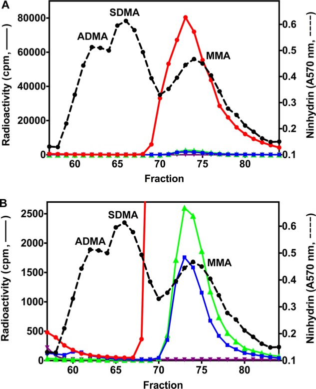
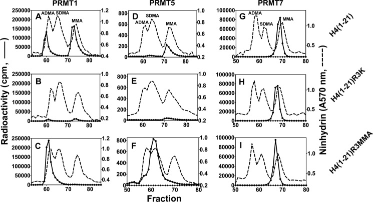
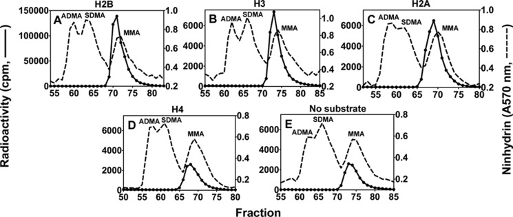
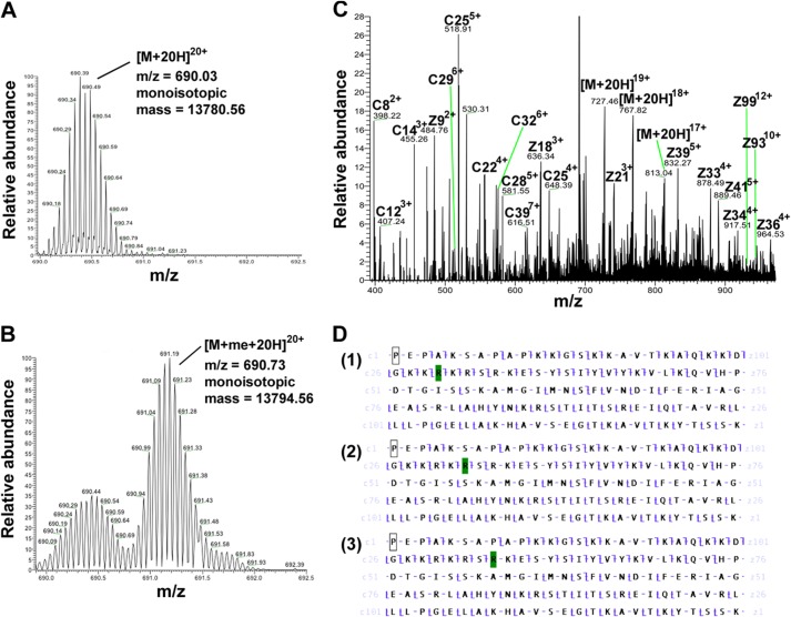
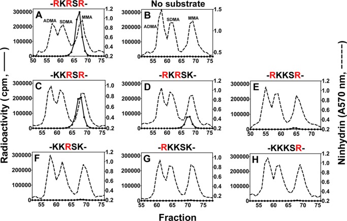
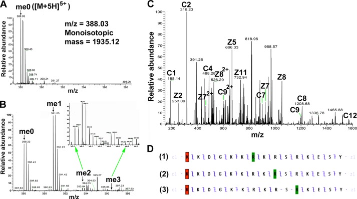
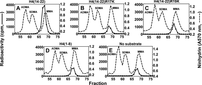
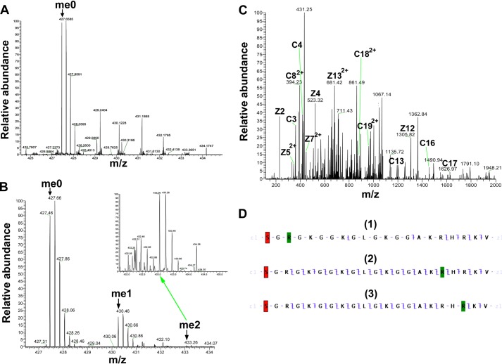
The mammalian protein arginine methyltransferase 7 (PRMT7) has been implicated in roles of transcriptional regulation, DNA damage repair, RNA splicing, cell differentiation, and metastasis. However, the type of reaction that it catalyzes and its substrate specificity remain controversial. In this study, we purified a recombinant mouse PRMT7 expressed in insect cells that demonstrates a robust methyltransferase activity. Using a variety of substrates, we demonstrate that the enzyme only catalyzes the formation of ω-monomethylarginine residues, and we confirm its activity as the prototype type III protein arginine methyltransferase. This enzyme is active on all recombinant human core histones, but histone H2B is a highly preferred substrate. Analysis of the specific methylation sites within intact histone H2B and within H2B and H4 peptides revealed novel post-translational modification sites and a unique specificity of PRMT7 for methylating arginine residues in lysine- and arginine-rich regions. We demonstrate that a prominent substrate recognition motif consists of a pair of arginine residues separated by one residue (RXR motif). These findings will significantly accelerate substrate profile analysis, biological function study, and inhibitor discovery for PRMT7.

Keywords: Histone Methylation; Mass Spectrometry (MS); Methyltransferases; Post-translational Modification; Protein Methylation; Protein-arginine Methyltransferases (PRMT); S-Adenosylmethionine (AdoMet).


Figures

FIGURE 1.
FIGURE 1.
Reactions catalyzed by type I, II, and III PRMTs. AdoHcy, S-adenosylhomocysteine.
FIGURE 2.
FIGURE 2.
Recombinant mouse PRMT7 expressed in Sf9 insect cells. A, SDS-PAGE followed by Coomassie Blue staining of the purified wild-type enzyme, revealing a single major polypeptide of ∼80 kDa (expected mass is 81.2 kDa). This protein includes an N-terminal extension of GPLGY and a C-terminal extension of ELALVPRGSSAHHHHHHHHHH. The lane of marker proteins is shown on the left with the polypeptide sizes. B, SDS-PAGE analysis of the PRMT7 inactive mutant with motif I residues LDIG changed to AAAA. C, LC-MS/MS analysis of tryptic peptides of wild-type PRMT7. Sequences were searched against both the S. frugiperda and the mouse subsets of the UniRef100 database as well as the tagged expected sequence of PRMT7. Sequence coverage of the recombinant PRMT7 (85%) is shown by underlining; those residues not present in the endogenous protein are highlighted in bold and italics; the LDIG motif that was mutated in the PRMT7 mutant is shown in bold.
FIGURE 3.
FIGURE 3.
Formation of [3H]MMA from incubation of PRMT7 with [3H]AdoMet and GST-GAR as a methyl-accepting substrate. A, in vitro methylation reactions were performed as described under “Experimental Procedures” using 6 μg of GST-GAR and 1.2 μg of wild-type or mutant PRMT7 at a final concentration of 0.26 μm. Incubations in the absence of PRMT7 or GST-GAR were used as controls. The reactions were allowed to proceed at room temperature for 20 h. After acid hydrolysis, the methylated amino acid derivatives were analyzed by high resolution cation exchange chromatography together with standards of ADMA, SDMA, and MMA as described under “Experimental Procedures.” 3H radioactivity (solid lines) and the ninhydrin color of the methylated arginine standards (dashed lines; elution positions indicated) were determined with liquid scintillation counting and 570 nm absorbance, respectively. Because of a tritium isotope effect (5), the [3H]methyl derivatives of ADMA, SDMA, and MMA elute on the high resolution cation exchange chromatography column 1–2 min earlier than the nonisotopically labeled standards. Red, wild-type PRMT7 with GST-GAR; green, wild-type PRMT7 alone; blue, mutant PRMT7 with GST-GAR; purple, GST-GAR alone. B, magnification of the radioactivity scale to show PRMT7 automethylation (green), the residual activity of mutant PRMT7 (blue), and the absence of ADMA and SDMA in the reaction products (red).
FIGURE 4.
FIGURE 4.
Comparison of the methylation products of PRMT1, -5, and -7 using synthetic peptides derived from the N terminus of human histone H4. Peptides (12.5 μm final concentration) were incubated with [3H]AdoMet and PRMT1 (0.45 μm final concentration), PRMT5 (0.72 μm final concentration), or PRMT7 (0.26 μm final concentration) as described under “Experimental Procedures” and the reactions were allowed to proceed at room temperature for 20 h. The reaction mixtures were then acid-hydrolyzed and analyzed for methylated arginine species as described under “Experimental Procedures.” A–C show PRMT1-catalyzed methylation products of peptides H4(1–21), H4(1–21)R3K, and H4(1–21)R3MMA (Table 1), respectively. D–F show PRMT5-catalyzed methylation products of these peptides. G–I show PRMT7-catalyzed methylation products of these peptides. The elution positions of the ADMA, SDMA, and MMA standards are specifically indicated in A, D, and G but are similar in all of the experiments.
FIGURE 5.
FIGURE 5.
PRMT7-catalyzed methylation of core histones with and without buffer change detected by fluorography. 4 μg of human recombinant histones H2A, H2B, H3.3, and H4, either in the supplied buffer or exchanged into the reaction buffer (see “Experimental Procedures”), or 4 μg of GST-GAR were mixed with PRMT7 (0.26 μm final concentration) and [3H]AdoMet and incubated at room temperature as described under “Experimental Procedures” for 20 h in a final volume of 40 μl. Samples were then mixed with SDS loading buffer, separated on 4–12% BisTris gel, and stained with Coomassie Blue (A). The protein substrate in each lane was labeled at the bottom of the graph. The expected position of molecular mass standards (Bio-Rad, broad range) is shown on the left in kDa. The fluoroimage was developed after 7 days (d) of film exposure (B) or 3 h of film exposure (C). As described under “Experimental Procedures,” the density of radioactive bands from the film shown in C (where the density was mostly in the linear range) was divided by the density of the Coomassie-stained polypeptide to obtain the specific radioactivity (D). Data were normalized against the specific radioactivity of H2B (with buffer change).
FIGURE 6.
FIGURE 6.
Amino acid analysis of histones methylated by PRMT7. 6 μg of recombinant human H2A, H2B, H3.3, or H4 was dialyzed against the reaction buffer and then incubated for 20 h with [3H]AdoMet and PRMT7 (0.26 μm final concentration), hydrolyzed to amino acids, and analyzed by high resolution cation exchange chromatography as described in Fig. 2 legend. A–D show the PRMT7-catalyzed methylation products of H2B, H3.3, H2A, and H4. E shows the control of PRMT7 automethylation in the absence of histone substrates.
FIGURE 7.
FIGURE 7.
Detection of PRMT7-formed monomethylarginine sites in histone H2B using top-down mass spectrometry. 15 μg of H2B was incubated with 4.8 μg of PRMT7 (1 μm) and 200 μm AdoMet in the reaction buffer at room temperature for 24 h in a final volume of 60 μl. The methylation products were desalted using an OMIX C18 ZipTip and directly introduced into an LTQ-Orbitrap mass spectrometer by nano-ESI as described previously (30). A, control mass spectrum of unreacted H2B is shown with ion isolation of the 20-charge species (m/z = 690.03, monoisotopic mass = 13,780.56 Da; calculated monoisotopic mass 13,780.54 Da). B, mass spectrum of methylated H2B from the incubation mixture described above is shown with ion isolation of the 20-charge species (m/z = 690.73, monoisotopic mass = 13,794.56 Da; calculated monoisotopic mass 13,794.56 Da). C, ETD tandem mass spectrum of the 20-charge precursor of methylated H2B. Unit resolution was achieved on all product ions by operating the instrument at 30,000 resolution (at 400 m/z). To maximize signal intensity for better sequence coverage in the dissociation experiment, the ion isolation experimental window was kept wide enough that there was some contamination of the desired molecular ion with unmethylated H2B. The c and z ions were assigned assuming the methyl group is on H2B R29, as in panel d-1. D, c and z ion coverage for H2B when a methyl group is assigned on Arg-29 (D-1), Arg-R31 (D-2), or Arg-33 (D-3). The identified c and z ions are shown mapped onto the primary sequence (c ions of N-terminal origin are indicated by the slash pointing up and to the left; c ions, of C-terminal origin are indicated with a slash pointing down and to the right). In D-1, the presence of C29 and Z97 ions indicates the methylation of Arg-29. In D-2, the presence of C31 and Z97 ions indicates the methylation of either Arg-29 or Arg-31. In D-3, the presence of C34 and Z97 ions indicates the methylation of Arg-29, Arg-31, or Arg-33. Residues highlighted in green represent monomethylarginine sites.
FIGURE 8.
FIGURE 8.
Amino acid analysis of peptides derived from residues 23–37 of histone H2B methylated by PRMT7. Peptides (12.5 μm final concentration; Table 1) were methylated by PRMT7 as described in Fig. 3 legend. A–H show PRMT7-catalyzed methylation products of H2B(23–37), no substrate, H2B(23–37)R29K, H2B(23–37)R33K, H2B(23–37)R31K, H2B(23–37)R29K,R33K, H2B(23–37)R31K,R33K, and H2B(23–37)R29K,R31K, respectively. Partial sequences containing the target arginine sites and their lysine mutations are indicated above the panels. The arginine methylation sites present in each peptide are highlighted in red.
FIGURE 9.
FIGURE 9.
Detection of PRMT7-formed monomethylarginine sites in a peptide corresponding to residues 23–37 of histone H2B by mass spectrometry. 24 μm H2B(23–37) was incubated with 4.8 μg of PRMT7 (1.87 μm) and AdoMet (182 μm) in reaction buffer in a final volume of 33 μl at room temperature for 24 h. The methylation products were desalted with OMIX C18 ZipTip and directly introduced into an LTQ-Orbitrap mass spectrometer by nano-ESI, as in Fig. 6. A, control mass spectrum showing the +5 charged state of unmethylated H2B(23–37) (me0; m/z = 388.03, monoisotopic mass = 1935.11 Da; calculated monoisotopic mass = 1935.12 Da). B, LTQ-Orbitrap spectrum showing the +5 charged state of PRMT7-methylated H2B(23–37). The species detected included the unmethylated (me0), the monomethylated (me1; m/z = 390.83, monoisotopic mass = 1949.13 Da; calculated monoisotopic mass = 1949.14 Da), the doubly methylated (me2; m/z = 393.63, monoisotopic mass = 1963.14 Da; calculated monoisotopic mass = 1963.15 Da), and the triply methylated (me3; m/z = 396.43, monoisotopic mass = 1977.15 Da; calculated monoisotopic mass = 1977. 17 Da). The inset shows a magnification of the region for doubly and triply methylated H2B(23–37). C, ETD tandem mass spectrum of the 5-charge precursor of monomethylated H2B(23–37). The peaks corresponding to me1 in B were isolated and fragmented with ETD. Unit resolution was achieved on all product ions by operating the instrument at 60,000 resolution (at 400 m/z). Note that me1 is a mixture of Arg-29-methylated, Arg-31-methylated, and Arg-33-methylated species. The c and z ions were assigned assuming the methyl group is on Arg-31, as in panel D-2. D, c and z ion coverage for H2B(23–37) peptide when a methyl group is assigned on Arg-29 (D-1), Arg-31 (D-2), or Arg-33 (D-3). In D-1, the presence of C7 and Z9 ions indicates the monomethylation of Arg-29. In D-2, the presence of C9 and Z7 ions indicates the monomethylation of Arg-31. In D-3, the Z5 and Z6 ions supporting monomethylation of Arg-33 were not found, probably because Arg-33 is methylated to a very low extent, as suggested by B me3. Acetylation of the N-terminal lysine residue is indicated by red shading; monomethylation of arginine residues is indicated by green shading.
FIGURE 10.
FIGURE 10.
Detection of PRMT7-formed monomethylarginine sites in a peptide corresponding to residues 23–37 of histone H2B with Arg-29 to Lys mutation by mass spectrometry. 24 μm H2B(23–37)R29K was incubated with 4.8 μg of PRMT7 (1.87 μm) and AdoMet (182 μm) in the reaction buffer in a final volume of 33 μl at room temperature for 24 h. The methylation products were desalted with OMIX C18 ZipTip and directly introduced into an LTQ-Orbitrap mass spectrometer by nano-ESI as described above. A, mass spectrum showing the +5 charged state of PRMT7-methylated H2B(23–37)R29K. The species detected included the unmethylated (me0; m/z = 382.43, monoisotopic mass = 1907.11 Da; calculated monoisotopic mass = 1907.11 Da), the monomethylated (me1; m/z = 385.23, monoisotopic mass = 1921.12 Da; calculated monoisotopic mass = 1921.13 Da), doubly methylated (me2; m/z = 388.03, monoisotopic mass = 1935.11 Da; calculated monoisotopic mass = 1935.15 Da). The inset shows a magnification of the region for doubly methylated H2B(23–37)R29K. B, ETD tandem mass spectrum of the 5-charge precursor of monomethylated H2B(23–37)R29K. The peaks corresponding to me1 in A were isolated and fragmented with ETD. The c and z ions were assigned assuming the methyl group is on Arg-31, as in C-1. C, c and z ion coverage for H2B(23–37)R29K when a methyl group is assigned on Arg-31 (B-1) or Arg-33 (B-2). In C-1, the presence of C9 and Z7 ions indicates the monomethylation of Arg-31. C-2, the Z5 and Z6 ions supporting monomethylation of R33 were not found, probably because Arg-33 is methylated to a very low extent, as suggested in A, me2. Acetylation of the N-terminal lysine residue is indicated by red shading; monomethylation of arginine residues is indicated by green shading.
FIGURE 11.
FIGURE 11.
Amino acid analysis of peptides derived from residues 14–22 of histone H4 methylated by PRMT7. Peptides (12.5 μm; Table 1) were incubated with PRMT7 (0.26 μm) and [3H]AdoMet (0.7 μm) as described under “Experimental Procedures,” and the reactions were allowed to proceed at room temperature for 20 h. The reaction mixtures were then acid-hydrolyzed and analyzed for methylated arginine species as described. A–D show PRMT7-catalyzed methylation products of H4(14–22), H4(14–22)R17K, H4(14–22)R19K, and H4(1–8), respectively. E shows the control of PRMT7 automethylation.
FIGURE 12.
FIGURE 12.
Detection of PRMT7-formed monomethylarginine sites in a peptide corresponding to residues 1–21 of histone H4 by mass spectrometry. 24 μm H4(1–21) was incubated with 4.8 μg of PRMT7 (1.87 μm) and AdoMet (182 μm) in the reaction buffer in a final volume of 33 μl at room temperature for 24 h. The methylation products were desalted with OMIX C18 ZipTip and directly introduced into an LTQ-Orbitrap mass spectrometer by nano-ESI as described above. A, control mass spectrum showing the +5 charged state of unmethylated H4(1–21) (me0; m/z = 427.46, monoisotopic mass = 2132.25 Da; calculated monoisotopic mass = 2132.26 Da). B, mass spectrum showing the +5 charged state of PRMT7-methylated H4(1–21). The species detected included the unmethylated (me0), the monomethylated (me1; m/z = 430.26, monoisotopic mass = 2146.27 Da; calculated monoisotopic mass = 2146.27 Da), the doubly methylated (me2; m/z = 433.06, monoisotopic mass = 2160.26 Da; calculated monoisotopic mass = 2160.29 Da). The inset shows a magnification of the region for doubly methylated H4(1–21). C, ETD tandem mass spectrum of the 5-charge precursor of monomethylated H4(1–21). The peaks corresponding to me1 in B were isolated and fragmented with ETD. The c and z ions were assigned assuming the methyl group is on Arg-17, as in D-2. D, c and z ion coverage for H4(1–21) when a methyl group is assigned on Arg-3 (D-1), Arg-17 (D-2), or Arg-19 (D-3). D-1, the absence of majority of ions indicates that H4R3 is not a good methylation site for PRMT7. D-2, the presence of C17 and Z5 ions indicates monomethylation of Arg-17. D-3, the presence of C19 and Z5 ions indicates monomethylation of Arg-17 or Arg-19. Acetylation of the N-terminal lysine residue is indicated by red shading; monomethylation of arginine residues is indicated by green shading.
FIGURE 13.
FIGURE 13.
Crystal structure of H2B N-terminal basic regions in a nucleosome core particle (Protein Data Bank code 1KX5, 1.9-Å resolution). A, two molecules of histone H2B.2 (Xenopus laevis; cyan) from a histone octamer wrapped by two strands of nucleosomal DNA (Homo sapiens; orange). The H2B repression region (HBR; sequence, KKDGKKRRKTRK; residues highlighted as blue sticks) is at the root of the flexible N-terminal tail and exits the nucleosome through a minor groove channel formed by two DNA strands (1). B and C exhibit magnifications of HBR1 and HBR2 areas from A. They are both tightly associated with surrounding DNA. Noncovalent bonds with distance <5 Å are shown. Green, Gly-101 and Gly-102 are from histone H4.

Similar articles

Cited by

References

    1. Walsh C. T. (2006) Post-translational Modification of Proteins: Expanding Nature's Inventory, pp. 1–490, Roberts and Co. Publishers, Englewood, CO
    1. Bedford M. T., Clarke S. G. (2009) Protein arginine methylation in mammals: who, what, and why. Mol. Cell 33, 1–13 - PMC - PubMed
    1. Yang Y., Bedford M. T. (2013) Protein-arginine methyltransferases and cancer. Nat. Rev. Cancer 13, 37–50 - PubMed
    1. Miranda T. B., Miranda M., Frankel A., Clarke S. (2004) PRMT7 is a member of the protein arginine methyltransferase family with a distinct substrate specificity. J. Biol. Chem. 279, 22902–22907 - PubMed
    1. Zurita-Lopez C. I., Sandberg T., Kelly R., Clarke S. G. (2012) Human protein arginine methyltransferase 7 (PRMT7) is a type III enzyme forming ω-NG-monomethylated arginine residues. J. Biol. Chem. 287, 7859–7870 - PMC - PubMed

Publication types

MeSH terms