. 2013 Nov-Dec;7(6):496-500.
doi: 10.4161/pri.27190. Epub 2013 Nov 18.

SAXS structural study of PrP(Sc) reveals ~11 nm diameter of basic double intertwined fibers

Affiliations

SAXS structural study of PrP(Sc) reveals ~11 nm diameter of basic double intertwined fibers

Heinz Amenitsch et al. Prion. 2013 Nov-Dec.

Abstract

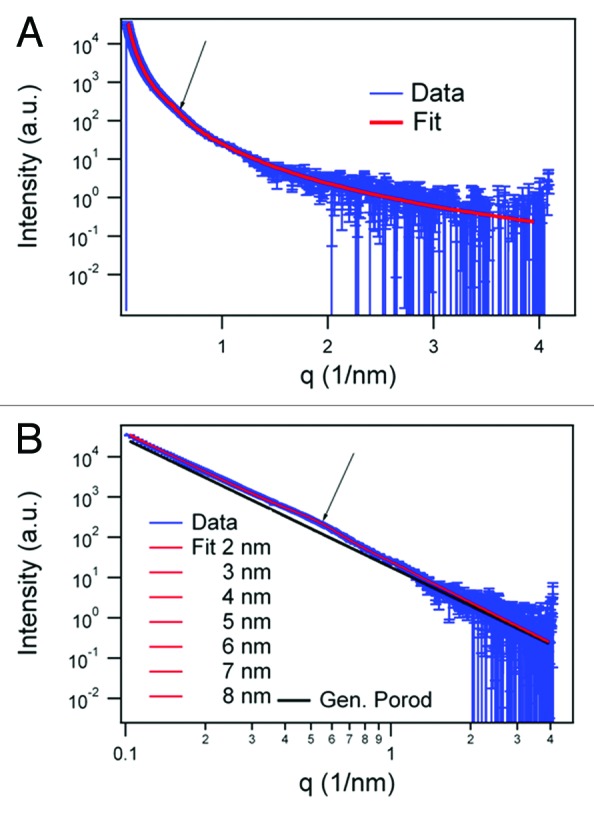
A sample of purified Syrian hamster PrP27-30 prion fibers was analyzed by synchrotron small-angle X-ray scattering (SAXS). The SAXS pattern obtained was fitted to a model based on infinitely long cylinders with a log-normal intensity distribution, a hard-sphere structure factor and a general Porod term for larger aggregates. The diameter calculated for the cylinders determined from the fit was 11.0 ± 0.2 nm. This measurement offers an estimation of the diameter of PrP(Sc) fibers in suspension, i.e., free of errors derived from estimations based on 2D projections in transmission electron microscopy images, subjected to further possible distortions from the negative stain. This diameter, which corresponds to a maximum diameter of approximately 5.5 nm for each of the two intertwined protofilaments making up the fibers, rules out the possibility that PrP(Sc) conforms to a stack of in-register, single-rung flat PrP(Sc) monomers; rather, PrP(Sc) subunits must necessarily coil, most likely several times, into themselves.

Keywords: PrPSc structure; SAXS; TEM; amyloid; prion; synchrotron radiation.


Figures

None
Figure 1. (A) Coomassie-stained SDS-PAGE of the SHaPrP27–30 sample subjected to SAXS analysis. (B) After SDS-PAGE, a sample was electroblotted onto a PVDF membrane and blotted with antibody 3F4. (C) Negative stain TEM of the same sample.
None
Figure 2. SAXS data in log-linear (A) representations and fit results of the infinite cylinder model (R = 5 nm) with hard-sphere structure factor and generalized Porod slope of SHaPrP27–30 (1 mg/mL). In the log-log representation of the SAXS data. (B) fit results varying the radius of the cylinders between 2 and 8 nm are shown together with the generalized Porod contribution (black). The arrow in both graphs indicates the hump of the structure factor corresponding to 11.0 ± 0.2 nm.
None
Figure 3. Schematic representation of PrPSc basic fibers based on the information obtained from SAXS and negative stain TEM.

References

    1. Prusiner SB. Prions. Proc Natl Acad Sci U S A. 1998;95:13363–83. doi: 10.1073/pnas.95.23.13363. - DOI - PMC - PubMed
    1. Safar J, Wille H, Itri V, Groth D, Serban H, Torchia M, Cohen FE, Prusiner SB. Eight prion strains have PrP(Sc) molecules with different conformations. Nat Med. 1998;4:1157–65. doi: 10.1038/2654. - DOI - PubMed
    1. Watson JD, Crick FH. Molecular structure of nucleic acids; a structure for deoxyribose nucleic acid. Nature. 1953;171:737–8. doi: 10.1038/171737a0. - DOI - PubMed
    1. Legname G, Baskakov IV, Nguyen HO, Riesner D, Cohen FE, DeArmond SJ, Prusiner SB. Synthetic mammalian prions. Science. 2004;305:673–6. doi: 10.1126/science.1100195. - DOI - PubMed
    1. Wang F, Wang X, Yuan CG, Ma J. Generating a prion with bacterially expressed recombinant prion protein. Science. 2010;327:1132–5. doi: 10.1126/science.1183748. - DOI - PMC - PubMed

Publication types

LinkOut - more resources