. 2014 Jan 1;74(1):153-161.
doi: 10.1158/0008-5472.CAN-13-1816. Epub 2013 Nov 18.

Inhibition of CSF-1 receptor improves the antitumor efficacy of adoptive cell transfer immunotherapy

Affiliations

Inhibition of CSF-1 receptor improves the antitumor efficacy of adoptive cell transfer immunotherapy

Stephen Mok et al. Cancer Res. .

Abstract

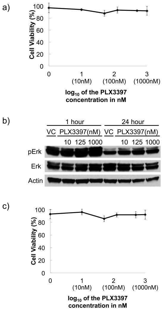
Colony stimulating factor 1 (CSF-1) recruits tumor-infiltrating myeloid cells (TIM) that suppress tumor immunity, including M2 macrophages and myeloid-derived suppressor cells (MDSC). The CSF-1 receptor (CSF-1R) is a tyrosine kinase that is targetable by small molecule inhibitors such as PLX3397. In this study, we used a syngeneic mouse model of BRAF(V600E)-driven melanoma to evaluate the ability of PLX3397 to improve the efficacy of adoptive cell therapy (ACT). In this model, we found that combined treatment produced superior antitumor responses compared with single treatments. In mice receiving the combined treatment, a dramatic reduction of TIMs and a skewing of MHCII(low) to MHCII(hi) macrophages were observed. Furthermore, mice receiving the combined treatment exhibited an increase in tumor-infiltrating lymphocytes (TIL) and T cells, as revealed by real-time imaging in vivo. In support of these observations, TILs from these mice released higher levels of IFN-γ. In conclusion, CSF-1R blockade with PLX3397 improved the efficacy of ACT immunotherapy by inhibiting the intratumoral accumulation of immunosuppressive macrophages.


Figures

Figure 1
Figure 1. Effects of PLX3397 on SM1 melanoma and primary T-cells
a) Murine SM1 melanoma cells were exposed to increasing concentrations of PLX3397 for 72 hours for IC50 determination using an MTS assay. b) Immunoblotting for analysis of signaling molecules after PLX3397 exposure of SM1 cells at 10, 125, 1000 nM for 1 or 24 hours. c) Effects of PLX3397 on murine splenocyte viability. Cell viability assay (MTS) of ex vivo activated C57BL/6 splenocytes at 72 hour time point with increasing doses of PLX3397.
Figure 2
Figure 2. Combined anti-tumor activity of adoptive cell transfer (ACT) immunotherapy and PLX3397 in the ovalbumin (OVA) and pmel-1 models
a) Schematic of the OT-1 ACT model based on adoptively transferring OT-1 splenocytes into lymphodepleted mice with previously established SM1 tumors stably expressing OVA antigen (SM1-OVA). b) Tumor growth curves of established SM1-OVA tumors in C57BL/6 mice through day 20 post-tumor implantation. c) Schematic of pmel-1 ACT model with lymphdepleted mice harboring SM1 tumors that adoptively received pmel-1 splenocytes and PLX3397. d) Tumor growth curves of established SM1 tumors in C57BL/6 mice through day 18.
Figure 3
Figure 3. Changes in intratumoral macrophages in responses to PLX3397
C57BL/6 mice with SM1-OVA tumors were gavaged p.o. daily with PLX3397 and received OT-1 ACT for 18 days to assess prolonged effects of the drug on macrophages. a) Tissue immunofluorescence microscopy to detect macrophages. Representative H&E (left) and immunofluorescence for macrophages stained with anti-F4/80-FITC (green, right), and nuclei stained with DAPI (blue, right). b) Cells stained for the surface expression markers of macrophages (F4/80+ CD11b+), M1-macrophage (MHCIIhi), and M2-macrophage (MHCIIlow) were used for FACS analysis. Bar-graph representation of percentage of F4/80(+) and CD11b(+) macrophages. c) Bar-graph representation of mean fluorescence intensity of MHCII expression on macrophages. d) Representative FACS plots demonstrating percentage of F4/80(+) CD11b(+) macrophages and mean fluorescence intensity of M1-marophage (MHCIIhi) and M2-macrophage (MHCIIlow) in tumor tissue.
Figure 4
Figure 4. Analysis of MDSC with PLX3397 exposure
C57BL/6 mice with SM1-OVA tumors were gavaged p.o. daily with PLX3397 and received OT-1 ACT for 18 days to assess prolonged effects of the drug on MDSC. a) Tissue immunofluorescence microscopy to detect MDSC on day 14 post-ACT. Representative H&E (left) and immunofluorescence for MDSC stained with anti-Gr-1-FITC (green, right), and nuclei stained with DAPI (blue, right). b) Cells stained for the surface expression markers of MDSC (Gr-1+ CD11b+), MO-MDSC (Gr-1low Ly6Chi), and PMN-MDSC (Gr-1hi Ly6Clow) were used for FACS analysis. Bar-graph representation of percentage of Gr-1(+) CD11b(+) MDSC. c) Bar-graph representation of ratio between MO-MDSC and PMN-MDSC. d) Representative FACS plots demonstrating percentages of Gr-1(+) CD11b(+) MDSC, MO-MDSC (Gr-1low Ly6Chi), and PMN-MDSC (Gr-1hi Ly6Clow) in tumor tissue.
Figure 5
Figure 5. Effects of PLX3397 mediated by CSF-1R and macrophages
a) Tumor growth curves of established SM1 tumors in C57BL/6 mice that received pmel-1 ACT with PLX3397, and anti-CSF-1 antibody. Treatment of anti-CSF-1 or isotype antibody control was started at the same time with PLX3397 when the tumor diameter reached 3 mm. On day 15, between vehicle+pmel-1 and anti-CSF-1+pmel-1, p=0.000008; between anti-CSF-1+pmel-1 and PLX3397+pmel, p=0.00003; between PLX3397+pmel-1 and PLX3397+anti-CSF-1+pmel-1, p=0.1 b) Tumor growth curves of established SM1 tumors in C57BL/6 mice treated PLX3397 in combination with clodronate. Clodronate treatment was started at the same time with PLX3397 when the tumor diameter reached 3 mm.
Figure 6
Figure 6. Effects of PLX3397 on the distribution and cytokine-producing functions of adoptively transferred lymphocytes
a) In vivo bioluminescence imaging of TCR transgenic T-cell distribution. OT-1 transgenic T-cells were transduced with a retrovirus-firefly luciferase and used for ACT. Representative figure at day 5 depicting 4 replicate mice per group. b) Quantitation of bioluminescence imaging of serial images with region of interest (ROI) analysis at the site of tumors obtained through day 13 post-ACT of OT-1 transgenic T-cells expressing firefly luciferase with 4 mice per group. c) Pmel-1 transgenic T-cells transduced with retrovirus-firefly luciferase were used for ACT. Day 5 representative figure showing 4 replicate mice per group. d) Quantitative measure of bioluminescence imaging signal with ROI at the tumor site obtained through day 10 post-ACT of luciferase expressing pmel-1 T-cells. e) Effects on cytokine production upon antigen restimulation. SM1 tumor-bearing C57BL/6 mice received pmel-1 ACT with or without PLX3397. At day 5 post-ACT, TILs were isolated for intracellular IFN-γ staining analyzed by FACS after 5 hour ex vivo exposure to the gp10025-33 peptide.
Figure 7
Figure 7. Lack of anti-tumor activity of PLX3397 in immunodeficient mice or with CD8+ T-cell depletion
a) Tumor growth curves of established SM1 tumors in non-irradiated C57BL/6 mice treated with PLX3397 in combination with anti-CD8 antibody. b) Tumor growth curves of established SM1 tumors in NSG mice treated with or without PLX3397. c) SM1 tumor-bearing mice received pmel-1 ACT and were treated with PLX3397 and anti-CD8 depleting antibody.

References

    1. Gabrilovich DI, Nagaraj S. Myeloid-derived suppressor cells as regulators of the immune system. Nat Rev Immunol. 2009;9:162–174. - PMC - PubMed
    1. Schreiber RD, Old LJ, Smyth MJ. Cancer immunoediting: integrating immunity's roles in cancer suppression and promotion. Science. 2011;331:1565–1570. - PubMed
    1. Kerkar SP, Restifo NP. Cellular constituents of immune escape within the tumor microenvironment. Cancer Res. 2012;72:3125–3130. - PMC - PubMed
    1. Priceman SJ, Sung JL, Shaposhnik Z, Burton JB, Torres-Collado AX, Moughon DL, Johnson M, Lusis AJ, Cohen DA, Iruela-Arispe ML, et al. Targeting distinct tumor-infiltrating myeloid cells by inhibiting CSF-1 receptor: combating tumor evasion of antiangiogenic therapy. Blood. 2010;115:1461–1471. - PMC - PubMed
    1. Tarhini AA, Butterfield LH, Shuai Y, Gooding WE, Kalinski P, Kirkwood JM. Differing patterns of circulating regulatory T cells and myeloid-derived suppressor cells in metastatic melanoma patients receiving anti-CTLA4 antibody and interferon-alpha or TLR-9 agonist and GM-CSF with peptide vaccination. J Immunother. 2012;35:702–710. - PMC - PubMed

Publication types

MeSH terms

Substances