. 2013 Dec 3;110(49):E4770-9.
doi: 10.1073/pnas.1309195110. Epub 2013 Nov 18.

Coxiella burnetii effector protein subverts clathrin-mediated vesicular trafficking for pathogen vacuole biogenesis

Affiliations

Coxiella burnetii effector protein subverts clathrin-mediated vesicular trafficking for pathogen vacuole biogenesis

Charles L Larson et al. Proc Natl Acad Sci U S A. .

Abstract

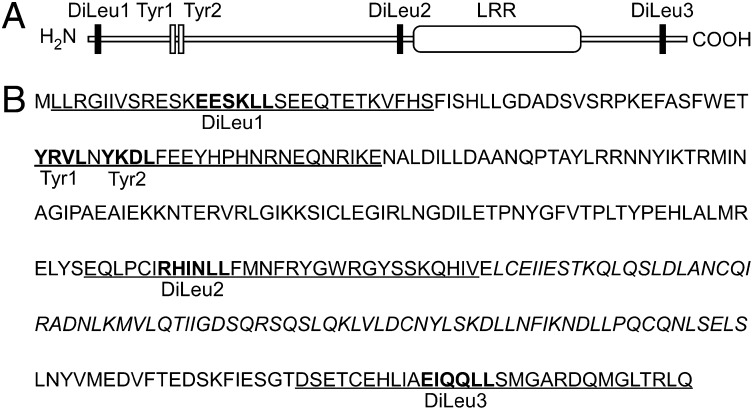
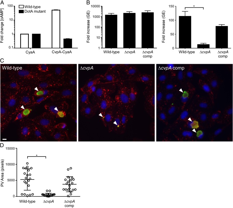
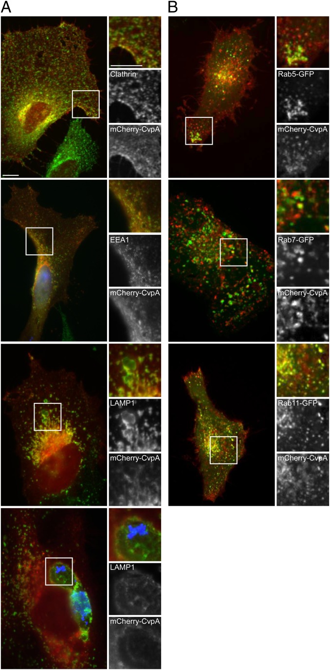
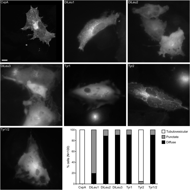
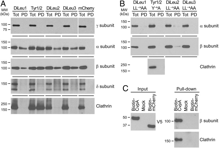
Successful macrophage colonization by Coxiella burnetii, the cause of human Q fever, requires pathogen-directed biogenesis of a large, growth-permissive parasitophorous vacuole (PV) with phagolysosomal characteristics. The vesicular trafficking pathways co-opted by C. burnetii for PV development are poorly defined; however, it is predicted that effector proteins delivered to the cytosol by a defective in organelle trafficking/intracellular multiplication (Dot/Icm) type 4B secretion system are required for membrane recruitment. Here, we describe involvement of clathrin-mediated vesicular trafficking in PV generation and the engagement of this pathway by the C. burnetii type 4B secretion system substrate Coxiella vacuolar protein A (CvpA). CvpA contains multiple dileucine [DERQ]XXXL[LI] and tyrosine (YXXΦ)-based endocytic sorting motifs like those recognized by the clathrin adaptor protein (AP) complexes AP1, AP2, and AP3. A C. burnetii ΔcvpA mutant exhibited significant defects in replication and PV development, confirming the importance of CvpA in infection. Ectopically expressed mCherry-CvpA localized to tubular and vesicular domains of pericentrosomal recycling endosomes positive for Rab11 and transferrin receptor, and CvpA membrane interactions were lost upon mutation of endocytic sorting motifs. Consistent with CvpA engagement of the endocytic recycling system, ectopic expression reduced uptake of transferrin. In pull-down assays, peptides containing CvpA-sorting motifs and full-length CvpA interacted with AP2 subunits and clathrin heavy chain. Furthermore, depletion of AP2 or clathrin by siRNA treatment significantly inhibited C. burnetii replication. Thus, our results reveal the importance of clathrin-coated vesicle trafficking in C. burnetii infection and define a role for CvpA in subverting these transport mechanisms.

Keywords: type IV secretion; vesicular fusion.


Conflict of interest statement

The authors declare no conflict of interest.

Figures

Fig. 1.
Fig. 1.
C. burnetii CvpA (CBU0665) contains a eukaryotic-like LRR domain and multiple endocytic sorting motifs. (A) Schematic of CvpA (328 aa, 38.0 kDa) showing the LRR and distribution of dileucine (DiLeu) and tyrosine (Tyr) endocytic sorting motifs identified using the Eukaryotic Linear Motif resource for functional sites in proteins (www.ELM.eu.org). (B) CvpA amino acid sequence. Consensus dileucine [DERQ]XXXL[LI] and tyrosine (YXXΦ; Φ, amino acid with a bulky hydrophobic side chain) endocytic sorting motifs are shown in boldface type. The amino acid sequence of peptides used in GST-30mer pull-down assays are underlined, and the LRR sequence is italicized.
Fig. 2.
Fig. 2.
CvpA is a Dot/Icm T4BSS substrate required for intracellular growth of C. burnetii. (A) Production of cytosolic cAMP by THP-1 cells infected with C. burnetii expressing CyaA-CvpA (Left). Cell lysates were collected 48 h post infection (pi), and fold increases in cAMP were determined relative to lysates from cells infected with C. burnetii expressing CyaA alone. Values are mean ± SEM of duplicate samples and are representative of three independent experiments. (B) Replication of wild-type C. burnetii, the ΔcvpA mutant, and the complemented mutant in ACCM-2 (Left) and THP-1 macrophages (Right). Fold increases in GEs at 5 d pi are depicted. Results are expressed as the means of two biological replicates representative of three independent experiments. Error bars indicate SE from the means, and an asterisk indicates a statistically significant difference (P < 0.05). (C) Representative epifluorescent micrographs of Vero cells infected with wild-type C. burnetii, the ΔcvpA mutant, or the complemented mutant. Vero cells were infected for 5 d and then immunostained for LAMP1 (red), Coxiella (green), and DNA (blue). Arrowheads denote PVs. (Scale bar, 10 μm.) (D) Size of PVs generated by wild-type C. burnetii, the ΔcvpA mutant, and the complemented mutant after 5 d growth in Vero cells as measured using ImageJ (n = 20). Error bars indicate SE from the means, and the asterisk indicates a statistically significant difference (P < 0.05).
Fig. 3.
Fig. 3.
Ectopically expressed mCherry-CvpA localizes to endocytic vesicles and traffics to pericentrosomal REs. (A) Representative micrographs of fixed HeLa cells expressing mCherry-CvpA (red) and immunostained for the vesicle proteins clathrin, EEA1, and LAMP1 (green). (Bottom) A cell infected with C. burnetii where bacteria and LAMP1 are immunostained blue and green, respectively. (B) Micrographs of live cells coexpressing mCherry-CvpA (red) and the GFP-tagged Rab GTPases Rab5, Rab7, or Rab11 (green). (Scale bar, 10 μm.)
Fig. 4.
Fig. 4.
Mutation of CvpA endocytic sorting motifs disrupts vesicular localization of mCherry-CvpA. Representative micrographs of HeLa cells expressing mCherry-CvpA or mCherry-CvpA proteins where diluecine residues were substituted with dialanine in DiLeu1, Dileu2, and Dileu3, and tyrosine residues were substituted with alanine in Tyr1, Tyr2, and Tyr1/Tyr2. (Scale bar, 10 μm.) (Lower Right) Graph showing the percentage of HeLa cells exhibiting tubulovesicular, punctate, or diffuse localization of ectopically expressed mCherry-CvpA proteins (n = 100).
Fig. 5.
Fig. 5.
CvpA endocytic sorting motifs interact with AP2 and clathrin. (A) Pull-down assays using HeLa cell lysates and GST fusions to 30mer peptides containing the CvpA endocytic sorting motifs DiLeu1 (aa 2–31), DiLeu2 (aa 172–201), DiLeu3 (aa 299–328), or Tyr1/Tyr2 (aa 52–81). GST peptides and interacting proteins were collected with glutathione agarose beads and analyzed by immunoblot with antibodies against clathrin and clathrin adaptor complex subunits present in AP1 (subunit γ), AP2 (subunits α and β), or AP3 (subunit δ). Pull-down with GST-mCherry was used as a negative control. (B) Pull-down assays using GST fusions to 30mer peptides containing endocytic sorting motifs where diluecine residues were substituted with dialanines, and tyrosine residues were substituted with alanine. Immunoblotting was conducted with antibodies against the α- and β-subunits of AP2 or clathrin (Tyr1/Tyr2 pull-down). (C) Pull-down assay using biotinylated full-length CvpA. CvpA containing an N-terminal biotinylation signal and a C-terminal V5 tag was expressed in HeLa cells and then collected from cell lysates using streptavidin beads. Cells expressing biotin-mCherry or nontransfected cells (Mock) were used as negative controls. Immunoblot detection of the V5 tag was used to assess equal expression of biotin-tagged proteins (Left). Pull-down samples were immunoblotted with antibodies recognizing clathrin and the β-subunit of AP2 and clathrin (Right).
Fig. 6.
Fig. 6.
Depletion of cellular AP2 or clathrin inhibits C. burnetii intracellular growth. (A) Protein expression in cells treated with targeting (+) or nontargeting (NT) (−) siRNA at 0, 1, 3, 4, or 5 d post infection (pi). HeLa cells were transfected with siRNA to deplete γ- and μ-subunits of the AP1 complex (AP1G1 and AP1M1 siRNA), β- and μ-subunits of AP2 (AP2B1 and AP2M1 siRNA), or clathrin (CLTC siRNA) and then infected with C. burnetii 2 d later (day 0 pi). Cell lysates were immunoblotted with antibodies against the AP1 γ-subunit, AP2 β-subunit, or clathrin to assess protein depletion and with antibody against GAPDH to confirm equal protein loading. (B) C. burnetii replication in HeLa cells depleted of AP1 subunits, AP2 subunits, or clathrin by siRNA. C. burnetii GEs at 1, 3, and 5 d pi were compared with GEs at day 0 pi to determine fold increases. Results are expressed as the means of two biological replicates and are representative of two independent experiments. Error bars indicate SE from the means, and an asterisk indicates a statistically significant difference (P < 0.05).

References

    1. Maurin M, Raoult D. Q fever. Clin Microbiol Rev. 1999;12(4):518–553. - PMC - PubMed
    1. Voth DE, Heinzen RA. Lounging in a lysosome: The intracellular lifestyle of Coxiella burnetii. Cell Microbiol. 2007;9(4):829–840. - PubMed
    1. Alix E, Mukherjee S, Roy CR. Subversion of membrane transport pathways by vacuolar pathogens. J Cell Biol. 2011;195(6):943–952. - PMC - PubMed
    1. Isberg RR, O’Connor TJ, Heidtman M. The Legionella pneumophila replication vacuole: Making a cosy niche inside host cells. Nat Rev Microbiol. 2009;7(1):13–24. - PMC - PubMed
    1. Campoy EM, Zoppino FC, Colombo MI. The early secretory pathway contributes to the growth of the Coxiella-replicative niche. Infect Immun. 2011;79(1):402–413. - PMC - PubMed

Publication types

MeSH terms

Substances

LinkOut - more resources