. 2014 Feb;34(3):348-61.
doi: 10.1128/MCB.00825-13. Epub 2013 Nov 18.

Grb2 promotes integrin-induced focal adhesion kinase (FAK) autophosphorylation and directs the phosphorylation of protein tyrosine phosphatase α by the Src-FAK kinase complex

Affiliations

Grb2 promotes integrin-induced focal adhesion kinase (FAK) autophosphorylation and directs the phosphorylation of protein tyrosine phosphatase α by the Src-FAK kinase complex

Suzanne Y S Cheng et al. Mol Cell Biol. 2014 Feb.

Abstract

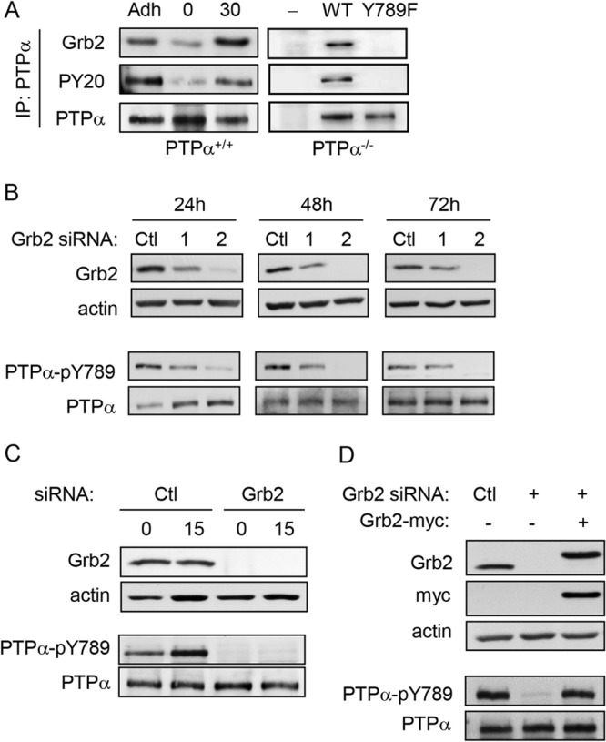
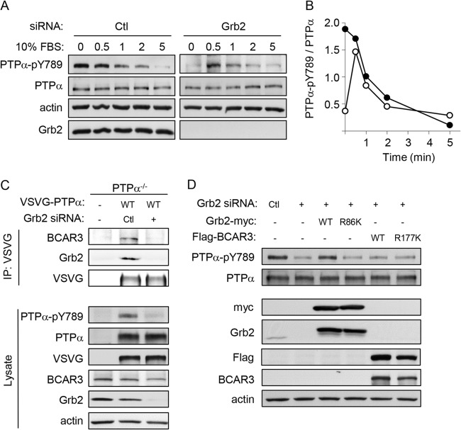
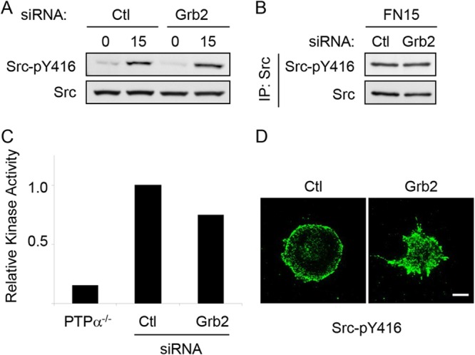
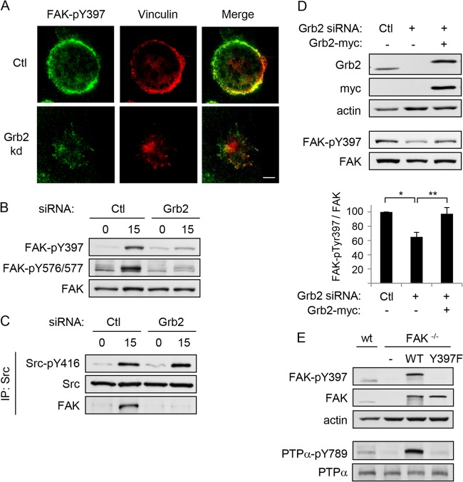
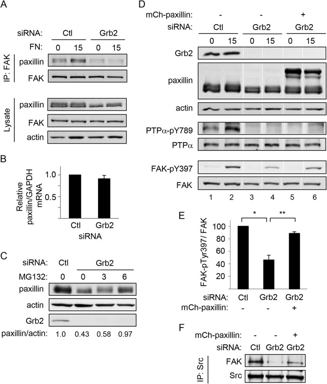
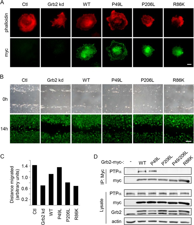
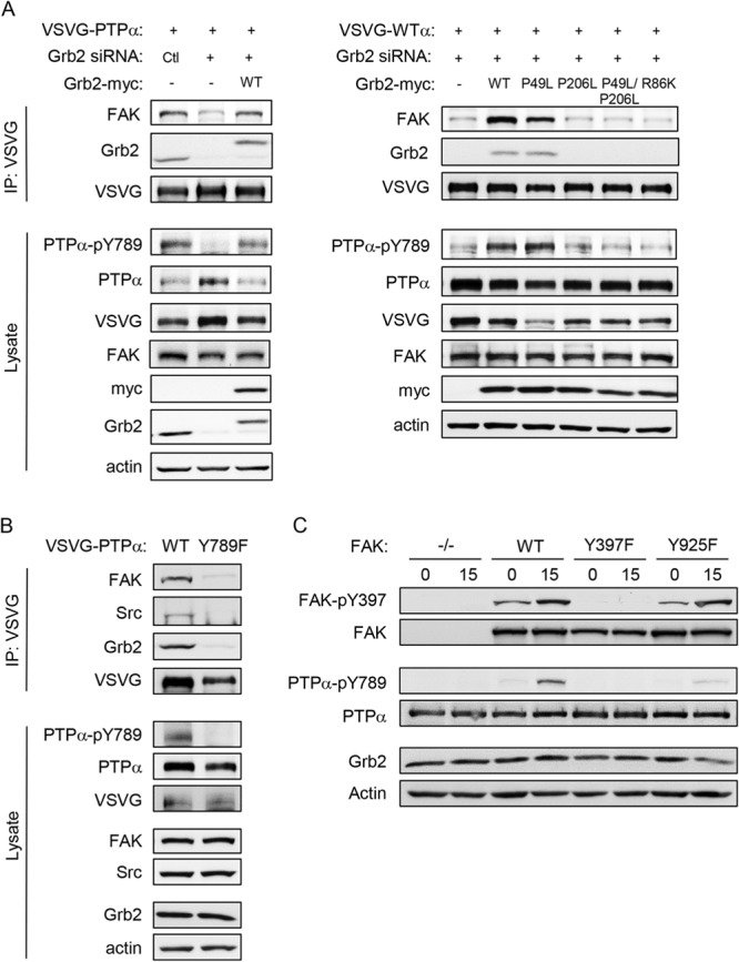
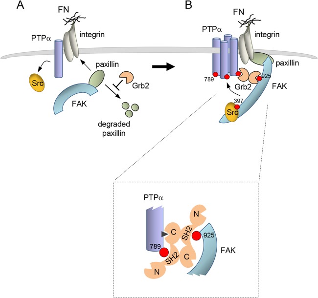
The integrin-activated Src-focal adhesion kinase (FAK) kinase complex phosphorylates PTPα at Tyr789, initiating PTPα-mediated signaling that promotes cell migration. Recruitment of the BCAR3-Cas complex by PTPα-phospho-Tyr789 at focal adhesions is one mechanism of PTPα signaling. The adaptor protein Grb2 is also recruited by PTPα-phospho-Tyr789, although the role of the PTPα-Grb2 complex in integrin signaling is unknown. We show that silencing Grb2 expression in fibroblasts abolishes PTPα-Tyr789 phosphorylation and that this is due to two unexpected actions of Grb2. First, Grb2 promotes integrin-induced autophosphorylation of FAK-Tyr397. This is impaired in Grb2-depleted cells and prohibits FAK activation and formation of the Src-FAK complex. Grb2-depleted cells contain less paxillin, and paxillin overexpression rescues FAK-Tyr397 phosphorylation, suggesting that the FAK-activating action of Grb2 involves paxillin. A second distinct role for Grb2 in PTPα-Tyr789 phosphorylation involves Grb2-mediated coupling of Src-FAK and PTPα. This requires two phosphosites, FAK-Tyr925 and PTPα-Tyr789, for Grb2-Src homology 2 (SH2) binding. We propose that a Grb2 dimer links FAK and PTPα, and this positions active Src-FAK in proximity with other, perhaps integrin-clustered, molecules of PTPα to enable maximal PTPα-Tyr789 phosphorylation. These findings identify Grb2 as a new FAK activator and reveal its essential role in coordinating PTPα tyrosine phosphorylation to enable downstream integrin signaling and migration.


Figures

FIG 1
FIG 1
Grb2 knockdown reduces PTPα Tyr789 phosphorylation. (A) Left panel, wild-type (PTPα+/+) MEFs were starved overnight (adherent cells [Adh]) and then trypsinized and kept in suspension for 1 h (0) before plating on FN-coated plates for 30 min (30). Right panel, PTPα-null MEFs (PTPα−/−) and PTPα-null MEFs reexpressing WT or Y789F-PTPα were stimulated on FN as described above. PTPα immunoprecipitates were probed for Grb2, phosphotyrosine (PY20), and PTPα. (B) Wild-type MEFs were treated with control (Ctl) or Grb2 siRNA duplex 1 or 2 for 24, 48, and 72 h. Cell lysates were probed for Grb2, actin, PTPα-pY789, and PTPα. (C) Wild-type MEFs were treated with control (Ctl) or Grb2 siRNA for 48 h and then starved overnight. Cells were trypsinized and held in suspension for 1 h (0), and then a portion of the suspended cells were plated on FN-coated plates for 15 min (15). Lysates were probed as shown. (D) Wild-type MEFs treated with either control (Ctl) or Grb2 siRNA or cotransfected with Grb2 siRNA and myc-tagged Grb2-WT were stimulated on FN-coated dishes for 15 min. Lysates were probed as shown.
FIG 2
FIG 2
Cell migration is reduced in the absence of Grb2. The migration of control (Ctl) and Grb2 siRNA-treated wild-type MEFs into the cleared area of a wounded cell monolayer was monitored for 24 h by time-lapse live-cell microscopy. Cell movement was tracked using ImageJ MtrackJ plugin. The two-dimensional (2D) random migration of 10 cells from each of the control (A) and Grb2 siRNA-treated (B) cell groups was measured and graphed. (C) The total distance of each track was measured for 10 cells in each group. The means are shown as horizontal bars and are significantly different (P = 0.001). (D) The net (linear) distance traveled was measured from the starting point of a track to the end. Results from 10 cell tracks are shown for each group. The means are shown as horizontal bars and are significantly different (P = 0.023).
FIG 3
FIG 3
PTPα-Tyr789 dephosphorylation and phosphorylation are not altered by expression of the phospho-Tyr789 binding proteins Grb2 and BCAR3. (A) Serum stimulation induces transient PTPα-Tyr789 phosphorylation in Grb2 knockdown cells. MEFs were treated with control or Grb2 siRNA, and 48 h later they were serum starved overnight and then stimulated with 10% FBS for 0.5, 1, 2, and 5 min. Lysates were immunoblotted with PTPα-pY789, PTPα, Grb2, and actin antibodies. (B) PTPα phosphorylation per unit of PTPα (arbitrary units) in control (Ctl) siRNA-treated (closed circles) and Grb2 siRNA-treated (open circles) cells was calculated from densitometric quantification of the blots shown in panel A. The absolute amount of PTPα Tyr789 phosphorylation cannot be compared between the control and Grb2 knockdown cells since a longer exposure of the autoradiograph from the latter lysate was quantified in order to accurately detect signal. (C) BCAR3 overexpression does not rescue phospho-PTPα-Tyr789. PTPα−/− MEFs were untreated or transfected with control (Ctl) or Grb2 siRNA and VSVG-tagged WT PTPα. Anti-VSVG immunoprecipitates from adherent cell lysates were probed for BCAR3, Grb2, and VSVG (VSVG-PTPα). Lysates were probed as shown. (D) Wild-type MEFs were transfected with either control (Ctl) or Grb2 siRNA or cotransfected with Grb2 siRNA and myc-Grb2-WT, myc-Grb2-R86K, Flag-BCAR3-WT, or Flag-BCAR3-R177K. Lysates were probed as shown.
FIG 4
FIG 4
Grb2 knockdown does not prevent integrin-induced Src activation. Wild-type MEFs treated with control (Ctl) or Grb2 siRNA were kept in suspension for 1 h (0) and replated on FN-coated coverslips or dishes for 15 min (15). Cell lysates (A) and Src immunoprecipitates from lysates of FN-stimulated cells (B) were probed for Src-phospho-Tyr416 (Src-pY416) and Src. (C) Src immunoprecipitates were prepared from lysates of PTPα−/− MEFs and from control (Ctl) and Grb2 siRNA-treated wild-type MEFs after FN stimulation for 15 min and used in in vitro kinase assays. Kinase activity is shown relative to that in the control cell immunoprecipitate (PTPα−/− cells, n = 1; control and Grb2 siRNA-treated cells, n = 2). (D) Cells plated on coverslips were washed, fixed, and immunostained with Src-pY416 antibody. Bar, 20 μm.
FIG 5
FIG 5
Integrin-induced FAK-Tyr397 phosphorylation and FAK/Src complex formation are dependent on Grb2. Wild-type MEFs treated with control (Ctl) or Grb2 siRNA were kept in suspension (0) for 1 h and replated on FN-coated coverslips/dishes for 15 min (15). (A) Cells on coverslips were washed, fixed, and immunostained with FAK-phospho-Tyr397 (FAK-pY397, green) and vinculin (red) antibodies. Bar, 20 μm. (B) Cell lysates were collected from the plates and probed for FAK-pY397, FAK-phospho-Y576/577 (FAK-pY576/577), and FAK. (C) Anti-Src immunoprecipitates were immunoblotted with Src-pY416, Src, and FAK. (D) MEFs transfected with either control (Ctl) or Grb2 siRNA or cotransfected with Grb2 siRNA and myc-tagged Grb2 were plated on FN-coated dishes for 15 min. Lysates were immunoblotted with Grb2, myc, actin, FAK-pY397, and FAK antibodies. The graph shows the amount of FAK-pTyr397 per unit of FAK calculated from 4 experiments, and the asterisks depict significant differences (*, P = 0.00003; **, P = 0.001). (E) Wild-type MEFs (wt), FAK-null MEFs (FAK−/−), and FAK-null MEFs stably reexpressing GFP-tagged wild-type (FAK-WT) or mutant (FAK-Y397F) FAK were immunoblotted with FAK-pY397, FAK, PTPα-pY789, PTPα, and actin antibodies.
FIG 6
FIG 6
Paxillin expression restores FAK activation but not PTPα-Ty789 phosphorylation in Grb2-depleted cells. MEFs treated with control (Ctl) or Grb2 siRNA were kept in suspension (0) for 1 h and replated on fibronectin (FN)-coated dishes for 15 min (15). (A) Immunoprecipitates of FAK were probed with FAK and paxillin (upper panels). Cell lysates were immunoblotted for FAK, paxillin, and actin (lower panels). (B) Relative levels of paxillin mRNA per unit of GAPDH mRNA in control (Ctl) and Grb2 siRNA-treated cells were determined by RT-PCR. (C) Untreated control (Ctl) and Grb2-depleted (0) cells and Grb2-depleted cells that were incubated with 20 μM MG132 for 3 and 6 h were probed for paxillin, actin, and Grb2. The bottom numbers show the relative units of paxillin per actin in each sample. (D) MEFs were treated with control (Ctl) or Grb2 siRNA or cotransfected with Grb2 siRNA and mCherry (mCh)-paxillin for 48 h and then starved in reduced serum medium overnight. The next day, cells were trypsinized, kept in suspension (0) for 1 h, and replated on FN-coated dishes for 15 min (15). Lysates were immunoblotted with Grb2, paxillin, actin, PTPα-pY789, PTPα, FAK-pY397, and FAK antibodies. Lanes 1 to 6 in each panel are from a single gel. The white vertical lines indicate that the samples in lanes 3 and 5 were inadvertently exchanged on the original gel, and these lane images were spliced out and repositioned correctly in the final image shown here. (E) The relative amount of FAK-pY397 per unit of FAK in FN-stimulated lysates from at least 3 experiments as in panel D was quantified by densitometry and is shown in the graph. Asterisks indicate significant differences (*, P = 0.0003; **, P = 0.0008). (F) Src immunoprecipitates from FN-stimulated lysates from experiments as in panel B were probed for FAK and Src.
FIG 7
FIG 7
Differential abilities of Grb2 domain mutants to rescue paxillin expression, FAK autophosphorylation at Tyr397, and PTPα-Tyr789 phosphorylation. (A) Schematic diagram of siRNA-resistant myc-tagged Grb2-WT/mutants. Silent mutations were made in the depicted region targeted by the Grb2 siRNA. The closed circles depict the domain with the inactivating point mutation. (B to E) MEFs treated with either control (Ctl) or Grb2 siRNA duplex or cotransfected with Grb2 siRNA and Grb2-myc-WT or mutants were kept in suspension for 1 h and replated on FN-coated dishes or coverslips for 15 min. (B) Paxillin expression was analyzed by probing cell lysates, and the graph in panel C shows the relative amount of paxillin per unit of FAK as calculated from densitometric analysis of 3 experiments as in panel B. (D) FAK-Tyr397 and PTPα-Tyr789 phosphorylation was determined by probing cell lysates (D), and the graph in panel E shows the relative amount of FAK-phospho-Tyr397 per unit of FAK as calculated from densitometric analysis of 3 experiments as in panel D.
FIG 8
FIG 8
Grb2 SH2 and C-terminal SH3 domains are required for cell spreading and migration and PTPα-Grb2 association. (A) MEFs treated with either control (Ctl) or Grb2 siRNA duplex or cotransfected with Grb2 siRNA and Grb2-myc-WT or Grb2-myc mutants were kept in suspension for 1 h and replated on FN-coated dishes for 15 min. Cells were fixed and immunostained with phalloidin (red) and myc (green). Bar, 20 μm. (B) Cells were plated on FN-coated coverslips for 48 h. A wound was created (0 h), and the cells were allowed to migrate into the cleared area for 14 h prior to staining with 1 nM calcein AM in medium. Bar, 250 μm. (C) The distance migrated by cells in two independent experiments as in panel B was measured as described in Materials and Methods, and the average is shown in the graph. (D) MEFs were transfected with Grb2-myc-WT or mutants. Anti-myc immunoprecipitates from adherent cell lysates were probed for PTPα and myc. Lysates were immunoblotted as shown.
FIG 9
FIG 9
Grb2 is required for FAK association with PTPα. (A) PTPα−/− MEFs were untreated or transfected as shown with control (Ctl) or Grb2 siRNA, myc-tagged Grb2-WT or the indicated Grb2 mutants, and VSVG-WT PTPα. VSVG immunoprecipitates from adherent cell lysates and cell lysates were probed as shown. (B) PTPα−/− MEFs were transfected with VSVG-WT-PTPα and VSVG-Y789F-PTPα. VSVG immunoprecipitates from adherent cell lysates and cell lysates were probed as shown. (C) FAK-null MEFs (FAK−/−) and FAK-null MEFs stably reexpressing GFP-tagged wild-type (FAK-WT) or the indicated mutant forms of FAK (Y397F or Y925F) were kept in suspension for 1 h (lanes 0) and replated on fibronectin (FN)-coated dishes for 15 min (lanes 15). Cell lysates were probed for the indicated (phospho)proteins.
FIG 10
FIG 10
Two roles of Grb2 in early integrin signaling. (A) Integrin engagement stimulates PTPα-catalyzed dephosphorylation and activation of Src. In role 1, Grb2 (depicted as a dimer, but this is unclear), promotes FAK autophosphorylation at Tyr397, possibly by mediating FAK relocalization in a paxillin-dependent manner. Right, FAK autophosphorylation at Tyr397 recruits Src, with consequent Src-catalyzed phosphorylation of FAK at activation loop tyrosines (not shown) and at Tyr925. (B) In role 2, a Grb2 dimer coordinates Src-FAK and PTPα complexation via Grb2-SH2 binding to FAK-phospho-Tyr925 and to PTPα-phospho-Tyr789 (details shown in boxed inset). The latter is suggested to represent a small population of PTPα that undergoes initial phosphorylation through an unknown mechanism or has basal level phosphorylation at Tyr789. The Grb2-mediated FAK-PTPα linkage positions active Src-FAK in proximity to nearby clustered molecules of PTPα to enable their phosphorylation at Tyr789. Additional cycles of Grb2-mediated Src-FAK binding to newly phosphorylated PTPα may be utilized to achieve maximal PTPα phosphorylation. Overall, a localized concentration of PTPα-phospho-Tyr789 that acts to signal downstream is generated, for example (not shown), by recruiting the BCAR3-Cas complex (18).

Similar articles

Cited by

References

    1. Geiger B, Yamada KM. 2011. Molecular architecture and function of matrix adhesions. Cold Spring Harb. Perspect. Biol. 3:pii a005033. 10.1101/cshperspect.a005033 - DOI - PMC - PubMed
    1. Huttenlocher A, Horwitz AR. 2011. Integrins in cell migration. Cold Spring Harb. Perspect. Biol. 3:a005074. 10.1101/cshperspect.a005074 - DOI - PMC - PubMed
    1. Burridge K, Sastry SK, Sallee JL. 2006. Regulation of cell adhesion by protein-tyrosine phosphatases. I. Cell-matrix adhesion. J. Biol. Chem. 281:15593–15596. 10.1074/jbc.R500030200 - DOI - PubMed
    1. Zaidel-Bar R, Itzkovitz S, Ma'ayan A, Iyengar R, Geiger B. 2007. Functional atlas of the integrin adhesome. Nat. Cell Biol. 9:858–867. 10.1038/ncb0807-858 - DOI - PMC - PubMed
    1. Parsons JT. 2003. Focal adhesion kinase: the first ten years. J. Cell Sci. 116:1409–1416. 10.1242/jcs.00373 - DOI - PubMed

Publication types

MeSH terms