. 2014 Jan;20(1):103-14.
doi: 10.1261/rna.041442.113. Epub 2013 Nov 18.

Biophysical characterization of G-quadruplex forming FMR1 mRNA and of its interactions with different fragile X mental retardation protein isoforms

Biophysical characterization of G-quadruplex forming FMR1 mRNA and of its interactions with different fragile X mental retardation protein isoforms

Anna C Blice-Baum et al. RNA. 2014 Jan.

Abstract

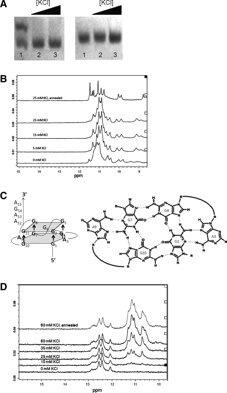
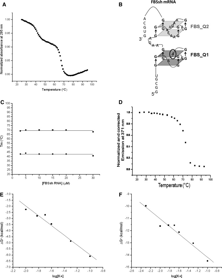
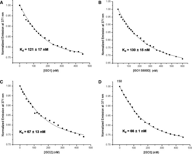
Fragile X syndrome, the most common form of inherited mental impairment in humans, is caused by the absence of the fragile X mental retardation protein (FMRP) due to a CGG trinucleotide repeat expansion in the 5'-untranslated region (UTR) and subsequent translational silencing of the fragile x mental retardation-1 (FMR1) gene. FMRP, which is proposed to be involved in the translational regulation of specific neuronal messenger RNA (mRNA) targets, contains an arginine-glycine-glycine (RGG) box RNA binding domain that has been shown to bind with high affinity to G-quadruplex forming mRNA structures. FMRP undergoes alternative splicing, and the binding of FMRP to a proposed G-quadruplex structure in the coding region of its mRNA (named FBS) has been proposed to affect the mRNA splicing events at exon 15. In this study, we used biophysical methods to directly demonstrate the folding of FMR1 FBS into a secondary structure that contains two specific G-quadruplexes and analyze its interactions with several FMRP isoforms. Our results show that minor splice isoforms, ISO2 and ISO3, created by the usage of the second and third acceptor sites at exon 15, bind with higher affinity to FBS than FMRP ISO1, which is created by the usage of the first acceptor site. FMRP ISO2 and ISO3 cannot undergo phosphorylation, an FMRP post-translational modification shown to modulate the protein translation regulation. Thus, their expression has to be tightly regulated, and this might be accomplished by a feedback mechanism involving the FMRP interactions with the G-quadruplex structures formed within FMR1 mRNA.

Keywords: FMRP; G-quadruplex RNA; fluorescence spectroscopy; protein–RNA interactions.


Figures

FIGURE 1.
FIGURE 1.
A schematic representation of the full-length FMRP, which shows the nuclear localization signal (NLS), the two K-homology domains (KH1 and KH2), the nuclear export signal (NES), the main site of phosphorylation (P), and the RGG box (RGG). An expansion of the sequence differences within isoforms 1–3, resulting from the alternative splicing at exon 15 of FMR1 mRNA, is also illustrated. The phosphorylation of serine 500 (arrow) has been shown to be biologically relevant. The bracket encompasses the amino acids encoded by the G-rich FBSsh region.
FIGURE 2.
FIGURE 2.
(A) Native PAGE of FBS_Q1 RNA (left) and FBS_Q2 RNA (right) in the presence of increasing KCl concentrations: 10 µM RNA at 0 mM KCl (lane 1), 25 mM KCl (lane 2), and 50 mM KCl (lane 3). The gels were visualized by UV shadowing at 254 nm. (B) 1D 1H-NMR spectra of 1.1 mM FBS_Q1 RNA in 10 mM cacodylic acid, pH 6.5, in the presence of increasing KCl concentrations in the range of 0–25 mM. (C) Proposed structure of FBS_Q2 (left); A:(G:G:G:G):A hexad formed by two adenines that are in the plane with the G-quartet (right). The formation of a hexad stabilizes the amino protons that are involved in Hoogsteen base-pairing within the G quartet. (D) 1D 1H-NMR spectra of 1.1 mM FBS_Q2 RNA in 10 mM cacodylic acid, pH 6.5, in the presence of increasing KCl concentrations in the range of 0–60 mM.
FIGURE 3.
FIGURE 3.
(A) Native PAGE of 30 µM FBS_Q1 RNA in 25 mM KCl in the presence of increasing concentrations of FMRP RGG box peptide: 0 µM RGG box (lane 1), 15 µM RGG box (lane 2), 30 µM RGG box (lane 3), and 60 µM RGG box (lane 4). (B) Native PAGE of 30 µM FBS_Q2 RNA in 25 mM KCl in the presence of increasing concentrations of FMRP RGG box peptide: 0 µM RGG box (lane 1), 15 µM RGG box (lane 2), 30 µM RGG box (lane 3), and 60 µM RGG box (lane 4). The gels were visualized by UV shadowing at 254 nm.
FIGURE 4.
FIGURE 4.
(A) 1D 1H-NMR spectra of 334 µM FBSsh RNA in 10 mM cacodylic acid, pH 6.5, in the presence of increasing KCl concentrations in the range of 0–25 mM. (B) Native PAGE of a fixed concentration of 10 µM FBSsh RNA in the presence of increasing concentrations of KCl in the range of 0–150 mM. (C) Native PAGE of increasing concentrations of FBSsh RNA in the range of 5–30 µM in the presence of 25 mM KCl. Gels were visualized by UV shadowing at 254 nm. (D) CD spectra of 10 µM FBSsh RNA in 10 mM cacodylic acid, pH 6.5, at 0 mM KCl, and 25 mM KCl.
FIGURE 5.
FIGURE 5.
(A) UV thermal denaturation curve of 10 µM FBSsh RNA in 10 mM cacodylic acid, pH 6.5, containing 25 mM KCl. (B) Model of FBSsh RNA showing the combination of the two G-quadruplexes formed by FBS_Q1 RNA and FBS_Q2 RNA. (C) Melting temperatures of both G-quadruplex structures of FBSsh RNA plotted against the RNA concentration. (D) Fluorescence spectroscopy thermal denaturation of 150 nM FBSsh_14AP RNA in 10 mM cacodylic acid, pH 6.5, and 25 mM KCl. (E) Plot of ΔG° as a function of the logarithm of K+ ion concentration for FBS_Q2. (F) Plot of ΔG° as a function of the logarithm of K+ ion concentration for FBS_Q1.
FIGURE 6.
FIGURE 6.
(A) FMRP ISO1 titrated into 150 nM FBSsh_14AP RNA in 10 mM cacodylic acid, pH 6.5, 750 nM BSA, and 25 mM KCl. (B) FMRP ISO1 S500D titrated into 150 nM FBSsh_14AP RNA in 10 mM cacodylic acid, pH 6.5, 750 nM BSA, and 25 mM KCl. (C) FMRP ISO2 titrated into 150 nM FBSsh_14AP RNA in 10 mM cacodylic acid, pH 6.5, 750 nM BSA, and 25 mM KCl. (D) FMRP ISO3 titrated into 150 nM FBSsh_14AP RNA in 10 mM cacodylic acid, pH 6.5, 750 nM BSA, and 25 mM KCl. Figures show a representative curve of the experiments performed in triplicate. The x-axis represents the total protein concentration [P]t from Equation 5 (Materials and Methods) for each FMRP isoform investigated.

References

    1. Akerman M, David-Eden H, Pinter RY, Mandel-Gutfreund Y 2009. A computational approach for genome-wide mapping of splicing factor binding sites. Genome Biol 10: 30 - PMC - PubMed
    1. Ashley CT, Sutcliffe JS, Kunst CB, Leiner HA, Eichler EE, Nelson DL, Warren ST 1993. Human and murine FMR-1: Alternative splicing and translational initiation downstream of the CGG-repeat. Nat Genet 4: 244–251 - PubMed
    1. Bharill S, Sarkar P, Ballin JD, Gryczynski I, Wilson GM, Gryczynski Z 2008. Fluorescence intensity decays of 2-aminopurine solutions: Lifetime distribution approach. Anal Biochem 377: 141–149 - PMC - PubMed
    1. Bole M, Menon L, Mihailescu MR 2008. Fragile X mental retardation protein recognition of G quadruplex structure per se is sufficient for high affinity binding to RNA. Mol Biosyst 4: 1212–1219 - PubMed
    1. Brackett DM, Qing F, Amieux PS, Sellers DL, Horner PJ, Morris DR 2013. Fmr1 transcript isoforms: Association with polyribosomes; regional and developmental expression in mouse brain. PLoS One 8: e58296. - PMC - PubMed

Publication types

MeSH terms

LinkOut - more resources