. 2014 Jan 14;129(2):157-72.
doi: 10.1161/CIRCULATIONAHA.113.006591. Epub 2013 Nov 18.

Growth properties of cardiac stem cells are a novel biomarker of patients' outcome after coronary bypass surgery

Affiliations

Growth properties of cardiac stem cells are a novel biomarker of patients' outcome after coronary bypass surgery

Domenico D'Amario et al. Circulation. .

Erratum in

  • Circulation. 2014 Aug 12;130(7):e65
  • Correction.
    [No authors listed] [No authors listed] Circulation. 2015 Dec 1;132(22):e363. doi: 10.1161/CIR.0000000000000341. Circulation. 2015. PMID: 26621666 No abstract available.

Retraction in

Expression of concern in

  • Expression of Concern.
    [No authors listed] [No authors listed] Circ Res. 2019 Jan 18;124(2):e4-e5. doi: 10.1161/RES.0000000000000241. Circ Res. 2019. PMID: 30582460 No abstract available.
  • Expression of Concern.
    [No authors listed] [No authors listed] Circulation. 2019 Jan 15;139(3):e5-e6. doi: 10.1161/CIR.0000000000000639. Circulation. 2019. PMID: 30615475 No abstract available.

Abstract

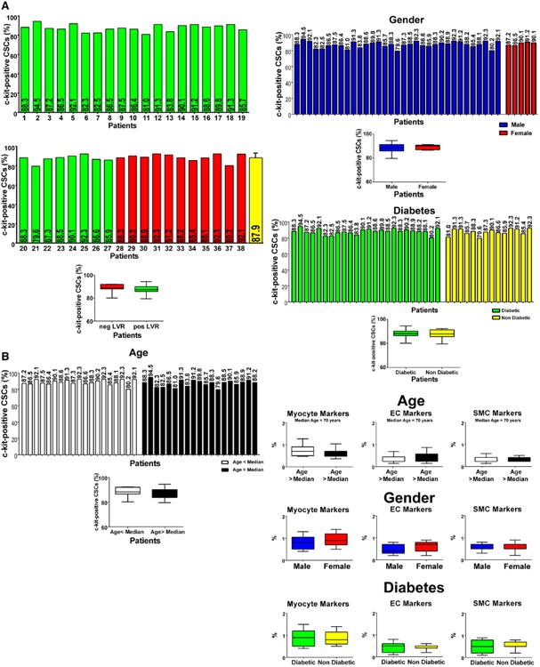
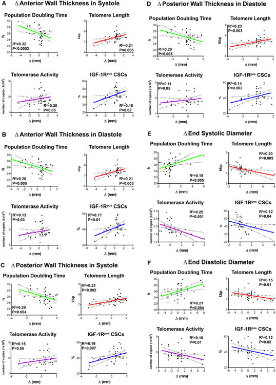
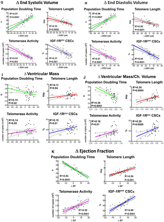
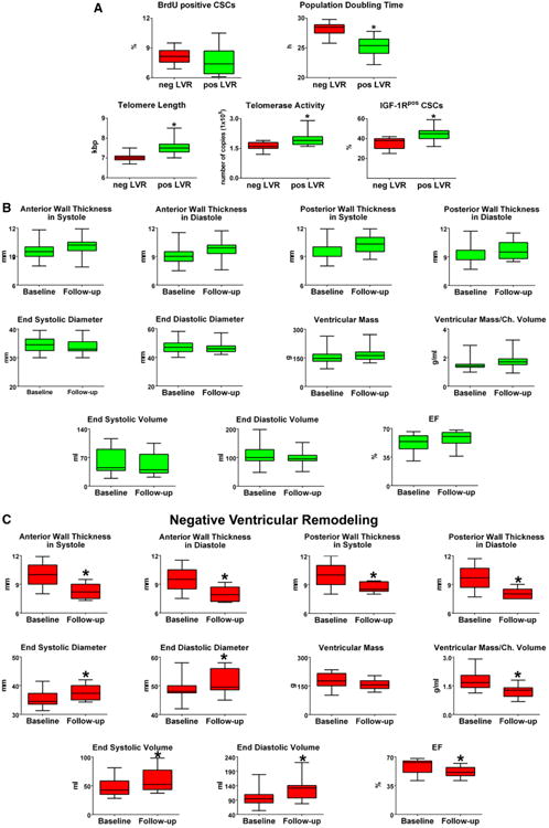
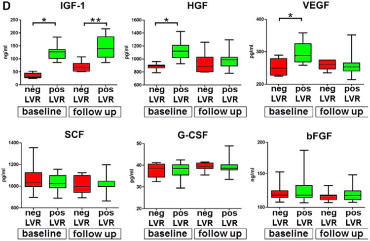
Background: The efficacy of bypass surgery in patients with ischemic cardiomyopathy is not easily predictable; preoperative clinical conditions may be similar, but the outcome may differ significantly. We hypothesized that the growth reserve of cardiac stem cells (CSCs) and circulating cytokines promoting CSC activation are critical determinants of ventricular remodeling in this patient population.

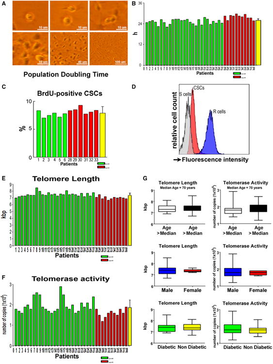
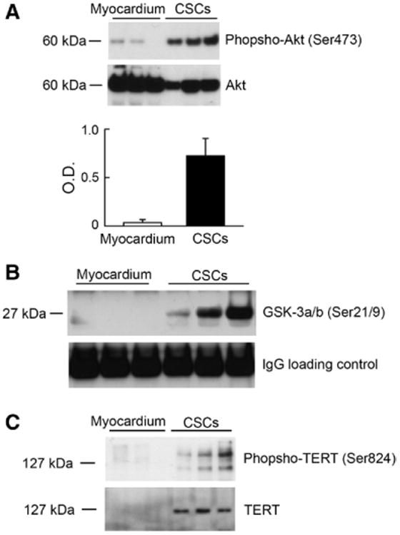
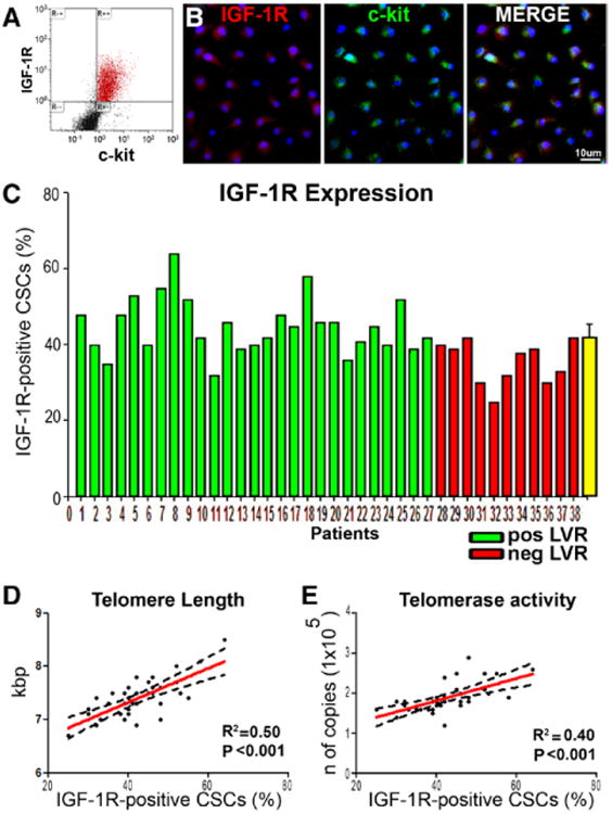
Methods and results: To document the growth kinetics of CSCs, population-doubling time, telomere length, telomerase activity, and insulin-like growth factor-1 receptor expression were measured in CSCs isolated from 38 patients undergoing bypass surgery. Additionally, the blood levels of insulin-like growth factor-1, hepatocyte growth factor, and vascular endothelial growth factor were evaluated. The variables of CSC growth were expressed as a function of the changes in wall thickness, chamber diameter and volume, ventricular mass-to-chamber volume ratio, and ejection fraction, before and 12 months after surgery. A high correlation was found between indices of CSC function and cardiac anatomy. Negative ventricular remodeling was not observed if CSCs retained a significant growth reserve. The high concentration of insulin-like growth factor-1 systemically pointed to the insulin-like growth factor-1-insulin-like growth factor-1 receptor system as a major player in the adaptive response of the myocardium. hepatocyte growth factor, a mediator of CSC migration, was also high in these patients preoperatively, as was vascular endothelial growth factor, possibly reflecting the vascular growth needed before bypass surgery. Conversely, a decline in CSC growth was coupled with wall thinning, chamber dilation, and depressed ejection fraction.

Conclusions: The telomere-telomerase axis, population-doubling time, and insulin-like growth factor-1 receptor expression in CSCs, together with a high circulating level of insulin-like growth factor-1, represent a novel biomarker able to predict the evolution of ischemic cardiomyopathy following revascularization.

Keywords: coronary artery disease; receptor, IGF type 1; stem cells; telomerase; telomere; ventricular remodeling.


Conflict of interest statement

Disclosures: None.

Figures

Figure 1
Figure 1
Phenotype of CSCs. A, Percentage of c-kit–positive CSCs in each of the 38 preparations. Patients 1 to 27 (color-coded green) experienced positive LVR; patients 28 to 38 (color-coded red) experienced negative LVR. B, Effects of age, sex, and diabetes mellitus on c-kit–positive CSCs and the expression of lineage markers. Data are shown by box plots: the box represents the interquartile range, the horizontal line inside the box marks the median, and whiskers show 5 to 95 percentiles range. CSC indicates cardiac stem cell; EC, endothelial cell; LVR, left ventricular remodeling; and SMC, smooth muscle cell.
Figure 2
Figure 2
Growth properties of CSCs. A, Phase-contrast images illustrating CSC division and growth in culture. B and C, Population-doubling time in the 38 CSC preparations (B), and bromodeoxyuridine (BrdU) labeling in 12 CSC preparations (C). D, Flow-FISH of CSCs and lymphoma cells of known telomere length (internal controls); CSCs were combined with lymphoma cells and incubated without (blank, not shown) or with PNA probe. The histogram represents the intensity of PNA probe binding in gated CSCs (green area), R cells (long telomeres: 48 kbp, blue area), and S cells (short telomeres: 7 kbp, grey area). Lymphoma cells were used to compute average telomere length in kbp. E, Telomere length in each of the 38 CSC preparations. F, Telomerase activity measured by qPCR in each of the 38 CSC samples. G, Effects of age, sex, and diabetes mellitus on telomere length and telomerase activity. Data are shown by box plots: the box represents the interquartile range, the horizontal line inside the box marks the median, and whiskers show 5 to 95 percentiles. CSC indicates cardiac stem cell; FISH, fluorescent in situ hybridization; PNA, peptide nucleic acid; and qPCR, quantitative polymerase chain reaction.
Figure 3
Figure 3
Akt and telomerase in CSCs. A, Phospho-Akt and total Akt were identified by Western blotting. Optical density (OD) data are shown. B, GSK-3α/β was used as a substrate for the detection of Akt kinase activity. Western blotting of phosphorylated GSK-3α/β at Ser21/9 is shown. Loading, IgG heavy chain band. C, Phosphotelomerase at Ser824 was detected following immunoprecipitation of CSC protein lysates with an antibody against telomerase. CSCs were obtained from patients 4, 6, 8, and 9, or 4, 6, and 9. CSC indicates cardiac stem cell; IgG, immunoglobulin G; and TERT, telomerase reverse transcriptase.
Figure 4
Figure 4
IGF-1R in CSCs. A, Bivariate distribution of c-kit and IGF-1R in CSCs. B, Confocal micrographs illustrating IGF-1R labeling (left, red) in c-kit–positive CSCs (center, green). Right, merge. C, Percentage of IGF-1R–positive CSCs measured by FACS in each of the 38 preparations. Patients 1 through 27 (color-coded green) experienced positive LVR; patients 28 through 38 (color-coded red) experienced negative LVR. D and E, Correlations between the percentage of IGF-1R–positive CSCs and telomere length (D), or telomerase activity (E). These relationships are shown by linear regression; solid lines represent the best-fit regression line associated with 95% confidence interval (dashed lines). P values represent the significance of the relationship expressed by the R2 values. Longer telomeres and higher telomerase activity correlate with positive LVR. CSC indicates cardiac stem cell; FACS, fluorescence-activated cell sorter; IGF-1R, insulin-like growth factor-1 receptor; and LVR, left ventricular remodeling.
Figure 5
Figure 5
Growth reserve of CSCs and anatomic indices of LVR. A through K, Correlations between PDT, telomere length, telomerase activity, and fraction of IGF-1R–positive (IGF-1Rpos) CSCs and the various parameters of cardiac size, shape, and function. These relationships are shown by linear regression; solid lines represent the best-fit regression line associated with 95% confidence interval (dashed lines). P values represent the significance of the relationship expressed by the R2 values. Ch. indicates left ventricular chamber; CSC, cardiac stem cell; EDV, end-diastolic volume; ESV, end-systolic volume; IGF-1R, insulin-like growth factor-1 receptor; LVR, left ventricular remodeling; and PDT, population-doubling time.
Figure 5
Figure 5
Growth reserve of CSCs and anatomic indices of LVR. A through K, Correlations between PDT, telomere length, telomerase activity, and fraction of IGF-1R–positive (IGF-1Rpos) CSCs and the various parameters of cardiac size, shape, and function. These relationships are shown by linear regression; solid lines represent the best-fit regression line associated with 95% confidence interval (dashed lines). P values represent the significance of the relationship expressed by the R2 values. Ch. indicates left ventricular chamber; CSC, cardiac stem cell; EDV, end-diastolic volume; ESV, end-systolic volume; IGF-1R, insulin-like growth factor-1 receptor; LVR, left ventricular remodeling; and PDT, population-doubling time.
Figure 6
Figure 6
CSCs, growth factors, and LVR. A through C, Data are shown by box plots: the box represents the interquartile range, the horizontal line inside the box marks the median, and whiskers show 5 to 95 percentiles. *Indicates P<0.05 vs negative (neg) LV remodeling (A, LVR) or baseline (B and C). Shorter population-doubling time, longer telomeres, and indices of wall thickening, decreased chamber size, and increased myocardial mass were coupled with positive LVR. D, Serum levels of IGF-1, HGF, VEGF, SCF, G-CSF, and bFGF at baseline and follow-up. The increase in IGF-1 was associated with positive LVR. The quantity of each growth factor in patients with positive (pos) and negative (neg) LVR is shown as mean±SD. *P<0.05 vs baseline values in patients who underwent neg LVR at follow-up; **P<0.05 vs follow-up values in patients who underwent neg LVR. bFGF indicates basic fibroblast growth factor; BrdU, bromodeoxyuridine; CSC, cardiac stem cell; G-CSF, granulocyte-colony stimulating factor; HGF, hepatocyte growth factor; IGF-1, insulin-like growth factor-1; IGF-1R, insulin-like growth factor-1 receptor; LVR, left ventricular remodeling; SCF, stem cell factor; SD, standard deviation; and VEGF, vascular endothelial growth factor.
Figure 6
Figure 6
CSCs, growth factors, and LVR. A through C, Data are shown by box plots: the box represents the interquartile range, the horizontal line inside the box marks the median, and whiskers show 5 to 95 percentiles. *Indicates P<0.05 vs negative (neg) LV remodeling (A, LVR) or baseline (B and C). Shorter population-doubling time, longer telomeres, and indices of wall thickening, decreased chamber size, and increased myocardial mass were coupled with positive LVR. D, Serum levels of IGF-1, HGF, VEGF, SCF, G-CSF, and bFGF at baseline and follow-up. The increase in IGF-1 was associated with positive LVR. The quantity of each growth factor in patients with positive (pos) and negative (neg) LVR is shown as mean±SD. *P<0.05 vs baseline values in patients who underwent neg LVR at follow-up; **P<0.05 vs follow-up values in patients who underwent neg LVR. bFGF indicates basic fibroblast growth factor; BrdU, bromodeoxyuridine; CSC, cardiac stem cell; G-CSF, granulocyte-colony stimulating factor; HGF, hepatocyte growth factor; IGF-1, insulin-like growth factor-1; IGF-1R, insulin-like growth factor-1 receptor; LVR, left ventricular remodeling; SCF, stem cell factor; SD, standard deviation; and VEGF, vascular endothelial growth factor.

Comment in

References

    1. Rizzello V, Poldermans D, Boersma E, Biagini E, Schinkel AF, Krenning B, Elhendy A, Vourvouri EC, Sozzi FB, Maat A, Crea F, Roelandt JR, Bax JJ. Opposite patterns of left ventricular remodeling after coronary revascularization in patients with ischemic cardiomyopathy: role of myocardial viability. Circulation. 2004;110:2383–2388. - PubMed
    1. Schinkel AF, Poldermans D, Rizzello V, Vanoverschelde JL, Elhendy A, Boersma E, Roelandt JR, Bax JJ. Why do patients with ischemic cardiomyopathy and a substantial amount of viable myocardium not always recover in function after revascularization? J Thorac Cardiovasc Surg. 2004;127:385–390. - PubMed
    1. Pfeffer MA, Braunwald E. Ventricular remodeling after myocardial infarction. Experimental observations and clinical implications. Circulation. 1990;81:1161–1172. - PubMed
    1. Pfeffer MA, Braunwald E, Moyé LA, Basta L, Brown EJ, Jr, Cuddy TE, Davis BR, Geltman EM, Goldman S, Flaker GC. Effect of captopril on mortality and morbidity in patients with left ventricular dysfunction after myocardial infarction. Results of the survival and ventricular enlargement trial. The SAVE Investigators. N Engl J Med. 1992;327:669–677. - PubMed
    1. Verma A, Meris A, Skali H, Ghali JK, Arnold JM, Bourgoun M, Velazquez EJ, McMurray JJ, Kober L, Pfeffer MA, Califf RM, Solomon SD. Prognostic implications of left ventricular mass and geometry following myocardial infarction: the VALIANT (VALsartan In Acute myocardial iNfarcTion) Echocardiographic Study. JACC Cardiovasc Imaging. 2008;1:582–591. - PubMed

Publication types

MeSH terms