

Idiopathic calcinosis cutis of the vulva
- PMID: 24249900
- PMCID: PMC3827520
- DOI: 10.4103/0019-5154.119960
Idiopathic calcinosis cutis of the vulva
Abstract
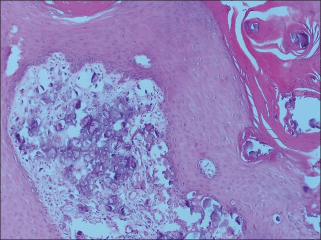
Background: In the present study, calcinosis cutis (CC) is defined as the deposition of amorphous calcium and phosphate salts under epidermis and it may be caused by a pre-existing event such as extravasation injury or hypercalcemic conditions. Idiopathic CC cases have no underlying disease or pre-existing cause.
Aim: A demostrative vulvar idiopathic CC case presentation and review of the related literature.
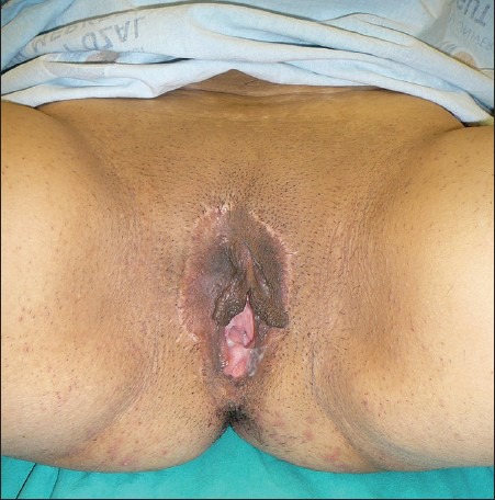
Methods: A 42-year-old multiparous female presented with vulvar nodular masses. She was keen on surgical removal of the lesions, as the masses caused dyscomfort during sexual intercourse. The lesions were removed and sent for histopathological examination. There was neither a history of trauma nor any inflammatory process in the vulvar skin prior to the development of lesions and no systemic abnormality was detected.
Results and conclusions: The histhopathologic evaluation of the biopsy specimen showed amorphous calcium deposits without any inflammatory infiltration in the dermis. There was no recurrence at 1 year's follow-up. This case shows that idiopathic CC may develop slowly at labio-vulvar region in a sexually active female with normal systemic or laboratory findings.
Keywords: Idiopathic calcinosis cutis; subepidermal nodules; vulva.
Conflict of interest statement
Figures


References
-
- Skidmore RA, Davis DA, Woosley JT, McCauliffe DP. Massive dystrophic calcinosis cutis secondary to chronic needle trauma. Cutis. 1997;60:259–62. - PubMed
-
- Ozçelik B, Serin IS, Başbuğ M, Oztürk F. Idiopathic calcinosis cutis of the vulva in an elderly woman. A case report. J Reprod Med. 2002;47:597–9. - PubMed
-
- Aksoy HM, Ozdemir R, Karaaslan O, Tiftikcioglu YO, Oruç M, Koçer U. Incidental idiopathic calcinosis cutis in a rhytidectomy patient. Dermatol Surg. 2004;30:1145–7. - PubMed
-
- Bernardo BD, Huettner PC, Merritt DF, Ratts VS. Idiopathic calcinosis cutis presenting as labial lesions in children: Report of two cases with literature review. J Pediatr Adolesc Gynecol. 1999;12:157–60. - PubMed
-
- Sengezer M, Türegün M, Tip GA. Calcinosis cutis requiring sculpturing in scleroderma. Plast Reconstr Surg. 1996;98:1319–21. - PubMed
LinkOut - more resources
Full Text Sources
Other Literature Sources