

Bullous systemic lupus erythematosus
- PMID: 24249912
- PMCID: PMC3827532
- DOI: 10.4103/0019-5154.119973
Bullous systemic lupus erythematosus
Abstract
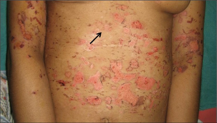
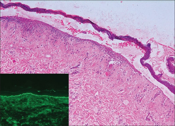
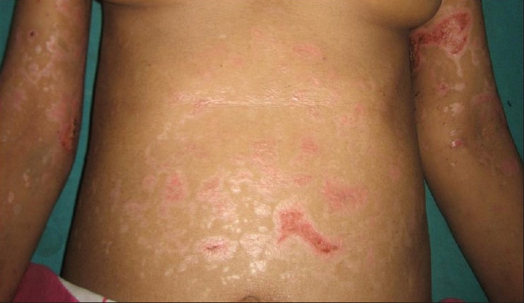
Bullous Systemic Lupus Erythematosus (BSLE) is a rare manifestation of Systemic Lupus Erythematosus (SLE), characterized histologically by subepidermal bullae with predominantly neutrophilic infiltrate and immunoglobulin (IgG, IgA, IgM) and C3 deposition at the basement membrane zone. Often associated is a heterogeneous profile of autoimmunity to components of type VII collagen. It needs to be differentiated from other subepidermal bullous diseases in view of potentially associated systemic manifestations of SLE. We report a 39-year-old lady who presented with an extensive vesicobullous eruption on face, neck, trunk, and mucosa. She was also found to have photosensitivity, significant anemia, and high antibody titers confirming SLE. Routine histopathology and immunoflorescence studies suggested BSLE and she responded favorably to dapsone. The importance of clinical and histopathological findings in confirming a diagnosis of BSLE along with the very encouraging response to dapsone therapy is highlighted.
Keywords: Dapsone; immunobullous diseases; type VII collagen.
Conflict of interest statement
Figures
Similar articles
-
Treatment of Bullous Systemic Lupus Erythematosus.J Immunol Res. 2015;2015:167064. doi: 10.1155/2015/167064. Epub 2015 May 18. J Immunol Res. 2015. PMID: 26090480 Free PMC article. Review.
-
Bullous systemic lupus erythematosus with lupus nephritis: a rare case of a subepidermal bullous disorder in a child.Cutis. 2012 Jan;89(1):17-21. Cutis. 2012. PMID: 22439305
-
Clinical, histological, immunological presentations and outcomes of bullous systemic lupus erythematosus: 10 New cases and a literature review of 118 cases.Semin Arthritis Rheum. 2018 Aug;48(1):83-89. doi: 10.1016/j.semarthrit.2017.11.003. Epub 2017 Nov 4. Semin Arthritis Rheum. 2018. PMID: 29191376
-
Bullous systemic lupus erythematosus: An uncommon manifestation in pediatric population.Andes Pediatr. 2021 Jun;92(3):428-433. doi: 10.32641/andespediatr.v92i3.2729. Andes Pediatr. 2021. PMID: 34479250 English, Spanish.
-
Bullous systemic lupus erythematosus.Australas J Dermatol. 2000 Nov;41(4):234-7. doi: 10.1046/j.1440-0960.2000.00426.x. Australas J Dermatol. 2000. PMID: 11105368 Review.
Cited by
-
A case of bullous systemic lupus erythematosus: Diagnostic challenges and clinical implications.Clin Case Rep. 2023 Nov 8;11(11):e8167. doi: 10.1002/ccr3.8167. eCollection 2023 Nov. Clin Case Rep. 2023. PMID: 37953897 Free PMC article.
-
Bullous Lupus: An Atypical Initial Presentation of Systemic Lupus Erythematosus Post-Delivery with Dramatic Response to Dapsone.Indian J Dermatol. 2022 Nov-Dec;67(6):763-765. doi: 10.4103/ijd.ijd_495_21. Indian J Dermatol. 2022. PMID: 36998892 Free PMC article. No abstract available.
-
Treatment of Bullous Systemic Lupus Erythematosus.J Immunol Res. 2015;2015:167064. doi: 10.1155/2015/167064. Epub 2015 May 18. J Immunol Res. 2015. PMID: 26090480 Free PMC article. Review.
-
Bullous lesions in a patient with systemic lupus erythematosus.J Clin Aesthet Dermatol. 2014 Sep;7(9):44-9. J Clin Aesthet Dermatol. 2014. PMID: 25276277 Free PMC article. Review.
-
Bullous systemic lupus erythematosus in a 10-year-old child.An Bras Dermatol. 2017;92(5 Suppl 1):37-39. doi: 10.1590/abd1806-4841.20176074. An Bras Dermatol. 2017. PMID: 29267441 Free PMC article.
References
-
- Yung A, Oakley A. Bullous systemic lupus erythematosus. Australas J Dermatol. 2000;41:234–7. - PubMed
-
- Chan LS, Lapiere JC, Chen M, Traczyk T, Mancini AJ, Paller AS, et al. Bullous systemic lupus erythematosus with antibodies recognizing multiple basement membrane components, bullous pemphigoid antigen1, lamini-5, laminin-6, and type VII collagen. Arch Dermatol. 1999;135:569–73. - PubMed
-
- Gammon WR, Briggaman RA. Bullous eruption of systemic lupus erythematosus. In: Wojnarowska F, Briggaman RA, editors. Management of Blistering Diseases. London: Chapman and Hall Ltd; 1990. pp. 263–75.
-
- Vassileve S. Bullous systemic lupus erythematosus. Clin Dermatol. 2004;22:129–38. - PubMed
-
- Gammon WR, Briggaman RA. Bullous SLE: A phenotypically distinctive but immunologically heterogeneous bullous disorder. J Invest Dermatol. 1993;100:28S–34. - PubMed
LinkOut - more resources
Full Text Sources
Other Literature Sources
Miscellaneous