

Bone scintigraphic patterns in patients of tumor induced osteomalacia
- PMID: 24250028
- PMCID: PMC3822419
- DOI: 10.4103/0972-3919.119541
Bone scintigraphic patterns in patients of tumor induced osteomalacia
Abstract
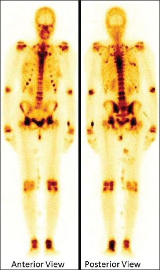
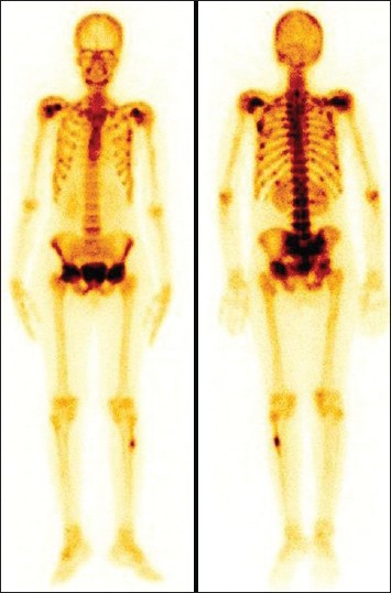
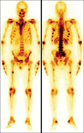
Tumor induced osteomalacia (TIO) or oncogenic osteomalacia is a rare condition associated with small tumor that secretes one of the phosphaturic hormones, i.e., fibroblast growth factor 23, resulting in abnormal phosphate metabolism. Patients may present with non-specific symptoms leading to delay in the diagnosis. Extensive skeletal involvement is frequently seen due to delay in the diagnosis and treatment. The small sized tumor and unexpected location make the identification of tumor difficult even after diagnosis of osteogenic osteomalacia. The bone scan done for the skeletal involvement may show the presence of metabolic features and the scan findings are a sensitive indicator of metabolic bone disorders. We present the bone scan findings in three patients diagnosed to have TIO.
Keywords: Bone scan; fibroblast growth factor 23; hypophosphatemia; tumor induced osteomalacia.
Conflict of interest statement
Figures
References
-
- Cai Q, Hodgson SF, Kao PC, Lennon VA, Klee GG, Zinsmiester AR, et al. Brief report: Inhibition of renal phosphate by a tumor product in a patient with oncogenic osteomalacia. N Engl J Med. 1994;330:1645–9. - PubMed
-
- Ryan EA, Reiss E. Oncogenous osteomalacia. Review of the world literature of 42 cases and report of two new cases. Am J Med. 1984;77:501–12. - PubMed
-
- Fukumoto S, Takeuchi Y, Nagano A, Fujita T. Diagnostic utility of magnetic resonance imaging skeletal survey in a patient with oncogenic osteomalacia. Bone. 1999;25:375–7. - PubMed
-
- Brame LA, White KE, Econs MJ. Renal phosphate wasting disorders: Clinical features and pathogenesis. Semin Nephrol. 2004;24:39–47. - PubMed
Publication types
LinkOut - more resources
Full Text Sources
Other Literature Sources
