

Malignant inflammatory myofibroblastic tumor of the maxillary sinus
- PMID: 24250100
- PMCID: PMC3830248
- DOI: 10.4103/0973-029X.119754
Malignant inflammatory myofibroblastic tumor of the maxillary sinus
Abstract
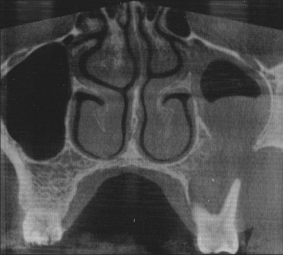
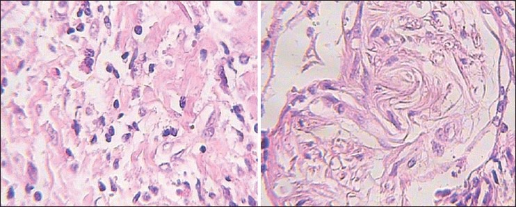
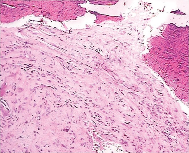
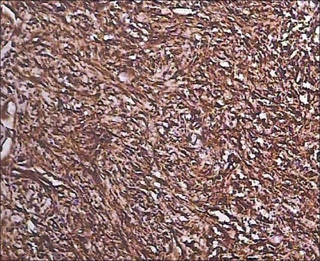
Inflammatory myofibroblastic tumors (IMTs) are extremely rare neoplasms with a variable natural history and biologic behavior, ranging from completely benign to malignant tumors with fatal outcome. They have no common identifiable cause, although some authors have assumed that any inflammatory stimulus may cause these pseudotumors. They are most commonly found in the lungs. Extrapulmonary sites include abdomen, retroperitoneum and extremities. IMTs rarely affect the head and neck, but the most common subsites in this region include the orbit, larynx, mouth, tonsils, parapharyngeal space, thyroid, parotid and lacrimal glands. There are few reports of inflammatory pseudotumors in the paranasal sinuses. In the maxillary sinus, the initial presenting sign is usually a nonspecific sinonasal mass, which has been growing over a period of weeks or months. On rare occasions, IMT may exhibit malignant transformation. Herein we present a rare case of pathologically proved IMT with malignant transformation which originated in the maxillary sinus of a 29-year-old male.
Keywords: Inflammatory myofibroblastic tumor; malignant transformation; maxillary sinus.
Conflict of interest statement
Figures
References
-
- Coffin CM, Hornick JL, Fletcher CD. Inflammatory myofibroblastic tumor: Comparison of clinicopathologic, histologic, and immunohistochemical features including ALK expression in atypical and aggressive cases. Am J Surg Pathol. 2007;31:509–20. - PubMed
-
- Fisher C. Myofibroblastic malignancies. Adv Anat Pathol. 2004;11:190–201. - PubMed
-
- Ushio M, Takeuchi N, Kikuchi S, Kaga K. Inflammatory pseudotumor of the paranasal sinuses: A case report. Auris Nasus Larynx. 2007;34:533–6. - PubMed
-
- Al-Sindi K, Al-Shehabi MH, Al-Khalifa SA. Inflammatory myofibroblastic tumor of paranasal sinuses. Saudi Med J. 2007;28:623–7. - PubMed
Publication types
LinkOut - more resources
Full Text Sources
Other Literature Sources
