

A serially coupled stationary phase method for the determination of urinary 8-oxo-7,8-dihydro-2'-deoxyguanosine by liquid chromatography ion trap tandem mass spectrometry
- PMID: 24251117
- PMCID: PMC3830056
- DOI: 10.1016/j.redox.2013.10.001
A serially coupled stationary phase method for the determination of urinary 8-oxo-7,8-dihydro-2'-deoxyguanosine by liquid chromatography ion trap tandem mass spectrometry
Abstract
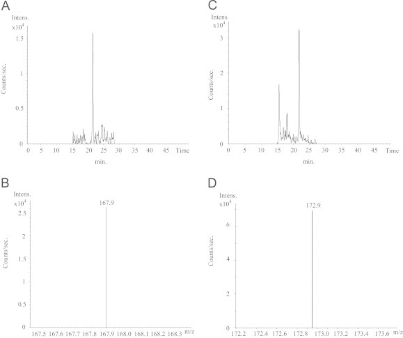
Oxidative attack to DNA is of particular interest since DNA modifications can lead to heritable mutations. The most studied product of DNA oxidation is 8-oxo-7,8-dihydro-2'-deoxyguanosine (8-oxodG). While 8-oxodG determination in blood and tissue cells is prone to artifacts, its measurement in urine employing liquid chromatography tandem mass spectrometry (LC-MS/MS) has gained more and more interest for increased reliability. LC-MS/MS can be affected by matrix effects and this is particularly true when ion trap is used as MS analyzer, due to ion accumulation in the trap and related space charge effect. In the present work, we have developed a LC-MS/MS method where the combination of cation exchange and reverse phase solid phases resulted in LC separation optimization. This together with the employment of an isotopically labeled internal standard, allowed the usage of ion trap LC-MS/MS, typically not employed for quantitative measurement in biological samples, for the measurement of 8-oxodG in urine samples from control populations. Four different urine matrices were employed for method validation. Limit of quantitation was set at least at 0.5 ng/ml. While analyzing urine samples from healthy volunteers, 8-oxodG levels reported as ng/ml were statistically different comparing males with females (p<0.05, Mann Whitney test); while comparing results normalized for creatinine no statistical significant difference was found. Mean urinary 8-oxodG level found in healthy volunteers was 1.16±0.46 nmol/mmol creatinine. The present method by enhancing at best the chromatographic performances allows the usage of ion trap LC-MS/MS for the measurement of 8-oxodG in urine samples from control populations.
Keywords: 15[N5]2-dG, 15[N5]2′-deoxyguanosine; 15[N5]8-oxodG, 8-oxo-7, 8-dihydro-15[N5]2′-deoxyguanosine; 8-oxo-7,8-dihydro-2′-deoxyguanosine; 8-oxodG, 8-oxo-7, 8-dihydro-2′-deoxyguanosine; EIC, Extracted Ion Chromatogram; ESI, electrospray ionization; IQC, internal quality control; IS, internal standard; Ion trap; LC-MS/MS; LC-MS/MS, liquid chromatography tandem mass spectrometry; LOQ, limit of quantitation; MRM, multiple reaction monitoring; MTH1, Nudix hydrolase mut T homologue 1; NER, nucleotide excision repair system; NIR, nucleotide incision repair system; Oxidative stress; ROS, reactive oxygen species; Reactive oxygen species; SACI, surface-activated ionization; TIC, Total Ion Chromatogram; Urine.
Figures


Similar articles
-
An improved liquid chromatography/tandem mass spectrometry method for the determination of 8-oxo-7,8-dihydro-2'-deoxyguanosine in DNA samples using immunoaffinity column purification.Rapid Commun Mass Spectrom. 2003;17(2):126-34. doi: 10.1002/rcm.883. Rapid Commun Mass Spectrom. 2003. PMID: 12512091
-
Rapid measurement of 8-oxo-7,8-dihydro-2'-deoxyguanosine in human biological matrices using ultra-high-performance liquid chromatography-tandem mass spectrometry.Free Radic Biol Med. 2012 May 15;52(10):2057-63. doi: 10.1016/j.freeradbiomed.2012.03.004. Epub 2012 Apr 15. Free Radic Biol Med. 2012. PMID: 22542794 Free PMC article.
-
Evaluation of enzyme-linked immunosorbent assay and liquid chromatography-tandem mass spectrometry methodology for the analysis of 8-oxo-7,8-dihydro-2'-deoxyguanosine in saliva and urine.Free Radic Biol Med. 2006 Dec 15;41(12):1829-36. doi: 10.1016/j.freeradbiomed.2006.09.009. Epub 2006 Sep 16. Free Radic Biol Med. 2006. PMID: 17157185
-
8-oxoguanine and 8-oxodeoxyguanosine Biomarkers of Oxidative DNA Damage: A Review on HPLC-ECD Determination.Molecules. 2022 Mar 1;27(5):1620. doi: 10.3390/molecules27051620. Molecules. 2022. PMID: 35268721 Free PMC article. Review.
-
The Intertwined Role of 8-oxodG and G4 in Transcription Regulation.Int J Mol Sci. 2023 Jan 19;24(3):2031. doi: 10.3390/ijms24032031. Int J Mol Sci. 2023. PMID: 36768357 Free PMC article. Review.
Cited by
-
Chemical Biomarkers of Exposure and Early Damage from Potentially Carcinogenic Airborne Pollutants.Ann Cancer Epidemiol. 2019 Sep;3:5. doi: 10.21037/ace.2019.08.01. Epub 2019 Sep 6. Ann Cancer Epidemiol. 2019. PMID: 40747480 Free PMC article.
-
Human Biomonitoring of DNA Adducts by Ion Trap Multistage Mass Spectrometry.Curr Protoc Nucleic Acid Chem. 2016 Sep 1;66:7.24.1-7.24.25. doi: 10.1002/cpnc.12. Curr Protoc Nucleic Acid Chem. 2016. PMID: 27584705 Free PMC article.
References
-
- Andreoli R., Manini P., De Palma G., Alinovi R., Goldoni M., Niessen W.M.A., Mutti A. Quantitative determination of urinary 8-oxo-7, 8-dihydro-2′-deoxyguanosine, 8-oxo-7, 8-dihydroguanine, 8-oxo-7, 8-dihydroguanosine, and their nonoxidized forms: daily concentration profile in healthy volunteers. Biomarkers. 2010;15:221–231. - PubMed
-
- Poulsen H.E., Nadal L.L., Broedbaek K., Nielsen P.E., Weimann A. Detection and interpretation of 8-oxodG and 8-oxoGua in urine, plasma and cerebrospinal fluid. Biochim. Biophys. Acta. 2013 〈http://dx.doi.org/10.1016/j.bbagen.2013.06.009〉 ([Epub ahead of print]) - DOI - PubMed
-
- Shibutani S., Takeshita M., Grollman A.P. Insertion of specific bases during DNA synthesis past the oxidation-damaged base 8-oxodG. Nature. 1991;349:431–434. - PubMed
-
- Collins A.R., Cadet J., Möller L., Poulsen H.E., Viña J. Are we sure we know how to measure 8-oxo-7,8-dihydroguanine in DNA from human cells? Arch. Biochem. Biophys. 2004;423:57–65. - PubMed
-
- Gedik C.M., Collins A. ESCODD, (European Standards Committee on Oxidative DNA Damage). Establishing the background level of base oxidation in human lymphocyte DNA: results of an interlaboratory validation study. FASEB J. 2005;19:82–84. - PubMed
MeSH terms
Substances
LinkOut - more resources
Full Text Sources
Other Literature Sources
Miscellaneous
