. 2013 Dec 3;52(48):8633-42.
doi: 10.1021/bi401325c. Epub 2013 Nov 19.

Bacterial toxin RelE: a highly efficient ribonuclease with exquisite substrate specificity using atypical catalytic residues

Affiliations

Bacterial toxin RelE: a highly efficient ribonuclease with exquisite substrate specificity using atypical catalytic residues

Meghan A Griffin et al. Biochemistry. .

Abstract

The toxin RelE is a ribosome-dependent endoribonuclease implicated in diverse cellular processes, including persistence. During amino acid starvation, RelE inhibits translation by cleaving ribosomal A-site mRNA. Although RelE is structurally similar to other microbial endoribonucleases, the active-site amino acid composition differs substantially and lacks obvious candidates for general acid-base functionality. Highly conserved RelE residues (Lys52, Lys54, Arg61, Arg81, and Tyr87) surround the mRNA scissile phosphate, and specific 16S rRNA contacts further contribute to substrate positioning. We used a single-turnover kinetic assay to evaluate the catalytic importance of individual residues in the RelE active site. Within the context of the ribosome, RelE rapidly cleaves A-site mRNA at a rate similar to those of traditional ribonucleases. Single-turnover rate constants decreased between 10(2)- and 10(6)-fold for the RelE active-site mutants of Lys52, Lys54, Arg61, and Arg81. RelE may principally promote catalysis via transition-state charge stabilization and leaving-group protonation, in addition to achieving in-line substrate positioning in cooperation with the ribosome. This kinetic analysis complements structural information to provide a foundation for understanding the molecular mechanism of this atypical endoribonuclease.


Figures

Figure 1
Figure 1. Structural insights into the RelE cleavage mechanism
The active site of (A) pre-cleavage and (B) post-cleavage co-crystal structures with RelE (purple) with a modified mRNA substrate (2′-O-methyl at nucleotide 20) or cyclic phosphate product (grey) and nearby ribosomal 16S rRNA (light green). Heteroatoms that may be important for catalysis are highlighted in blue (nitrogen) and red (oxygen). The arrow denotes the scissile phosphate, shown as an orange sphere. In the pre-cleavage structure, R81(*) is mutated to an alanine (PDB IDs: 3KIQ, 3KIU). (C) RelE induces a conformational change in the A-site mRNA. Overlay of mRNA in the 70S ribosomal A-site and P-site when RelE is bound (grey) or a cognate tRNA (not shown) is bound (orange) in the A-site. Dashed lines denote the corresponding mRNA nucleobases between each state (PDB ID: 2J00). (D) The structure of E.coli RelE from (A) (purple) superimposed on the structure of RNase T1 (green, PDB 1RGA).
Figure 2
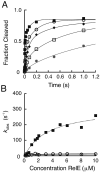
Figure 2. Wild-type RelE rapidly cleaves ribosomal RNA
(A) Measurement of single-turnover rates for wild-type RelE. Reactions contained 20 nM RC and increasing concentrations of wild-type RelE enzyme: 0.1 μM (*), 0.3 μM (□), 0.6 μM (◆), 1 μM (○), or 10 μM (■). The fractions of mRNA substrate cleaved during the first 1.2 seconds of each reaction is shown, but reactions were measured at least until 3 seconds and were fit as described in Experimental Procedures. (B) Dependence of cleavage rates on wild-type RelE enzyme concentration. The rates, kobs (s-1), for the fast (■) and slow phases (○) for a single replicate are plotted as a function of RelE concentration (μM). The rate constants (k2) and dissociation constants (Kd) were extracted from hyperbolic fits of each replicate and the mean and SEM for each constant is listed in Table 1 and Table S1.
Figure 3
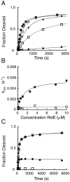
Figure 3. Analysis of single-turnover reaction kinetics of mutant RelE proteins
(A) Measurement of single-turnover rates for R81A RelE. Reactions contain 20 nM RC and increasing concentrations of R81A RelE enzyme: 0.1 μM (*), 0.3 μM (□), 0.6 μM (◆), 2.5 μM (○), or 10 μM (■). The first 3,000 seconds of the time course are shown and full time courses were fit as done for wild-type. (B) Dependence of cleavage rates on R81A RelE enzyme concentration. The rates, kobs (s-1), for the fast (■) and slow phases (○) for a single replicate are plotted as a function of RelE concentration (μM). The rate constants (k2) and dissociation constants (Kd) extracted from hyperbolic fits of each replicate and the mean and SEM for each constant is listed in Table 1 and Table S1. (C) Antitoxin RelB pulse-chase quench experiments with R81A RelE. Reactions contain 20 nM RC and 10 μM R81A RelE and were quenched either chemically (○), enzymatically with 6-fold excess RelB (■), or the RC was pre-mixed with RelB prior to addition of RelE (□). Product formation for the pulse-chase reaction with antitoxin RelB is plotted for the total time (reaction time, t1, + quench delay time or chase, t2) where RelB was added after t1 = 78 seconds (•).
Figure 4
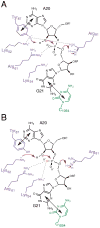
Figure 4. Mechanism of phosphodiester bond cleavage by RelE
(A) Previously proposed mechanism for RelE catalysis with RelE (purple), mRNA (black), and 16S rRNA C1054 (green) (12) . Tyr87 and C1054 stack with the second and third nucleotides, respectively (shown with black double arrows) to position the substrate for nucleophilic attack. The positively charged environment shifts the pKa of Tyr87, which could act a general base, while Arg61 stabilizes the transition state and Arg81 protonates the leaving group, acting as general acid. Dashed lines indicated proposed interactions. (B) Revised model for RelE catalysis, depicted as in (A). Here, Lys52 is activated as general base by the positively charged microenvironment, while Arg61 and Lys54 stabilize the transition state, and Arg81 acts as general acid. The large phosphorothioate effects suggest strong interactions between RelE and the pro-Sp non-bridging oxygen (orange), possibly from Arg61 and the neighboring Lys54.

Similar articles

Cited by

References

    1. Yamaguchi Y, Park JH, Inouye M. Toxin-antitoxin systems in bacteria and archaea. Annu Rev Genet. 2011;45:61–79. - PubMed
    1. Wang X, Wood TK. Toxin-antitoxin systems influence biofilm and persister cell formation and the general stress response. Appl Environ Microbiol. 2011;77:5577–5583. - PMC - PubMed
    1. Gerdes K, Maisonneuve E. Bacterial persistence and toxin-antitoxin loci. Annual review of microbiology. 2012;66:103–123. - PubMed
    1. Gerdes K. Toxin-antitoxin modules may regulate synthesis of macromolecules during nutritional stress. J Bacteriol. 2000;182:561–572. - PMC - PubMed
    1. Christensen SK, Mikkelsen M, Pedersen K, Gerdes K. RelE, a global inhibitor of translation, is activated during nutritional stress. Proc Natl Acad Sci U S A. 2001;98:14328–14333. - PMC - PubMed

Publication types

MeSH terms

LinkOut - more resources