. 2014 Feb;253(2):83-92.
doi: 10.1111/jmi.12099. Epub 2013 Nov 19.

Compact non-contact total emission detection for in vivo multiphoton excitation microscopy

Affiliations

Compact non-contact total emission detection for in vivo multiphoton excitation microscopy

Christian A Combs et al. J Microsc. 2014 Feb.

Abstract

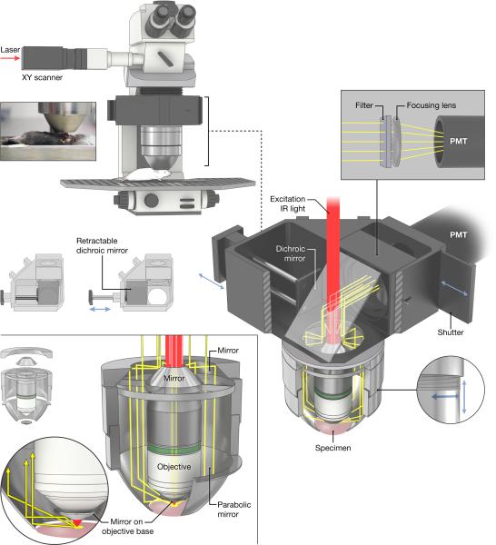
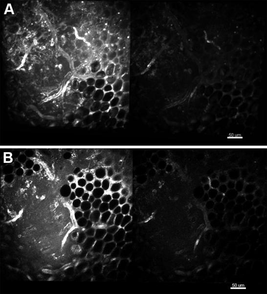
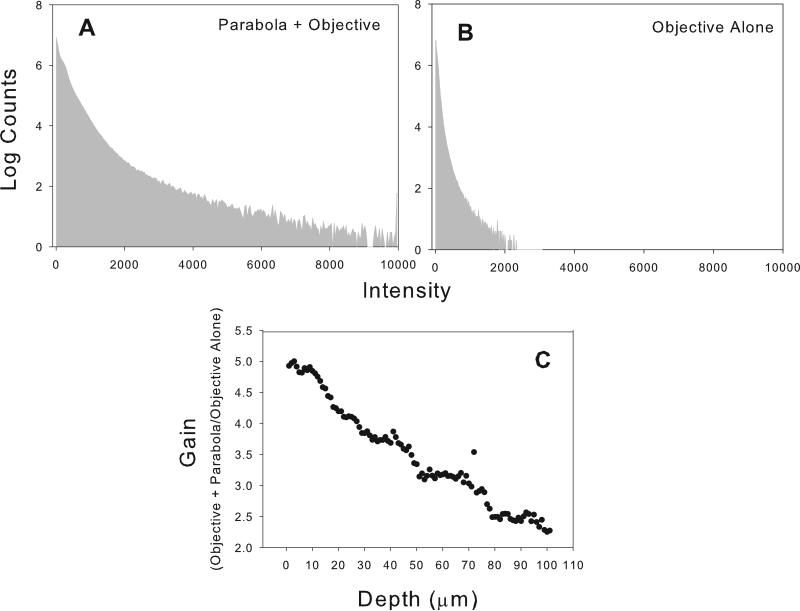
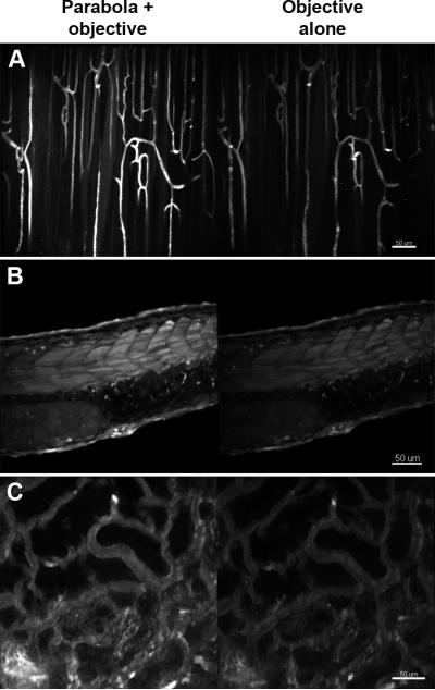
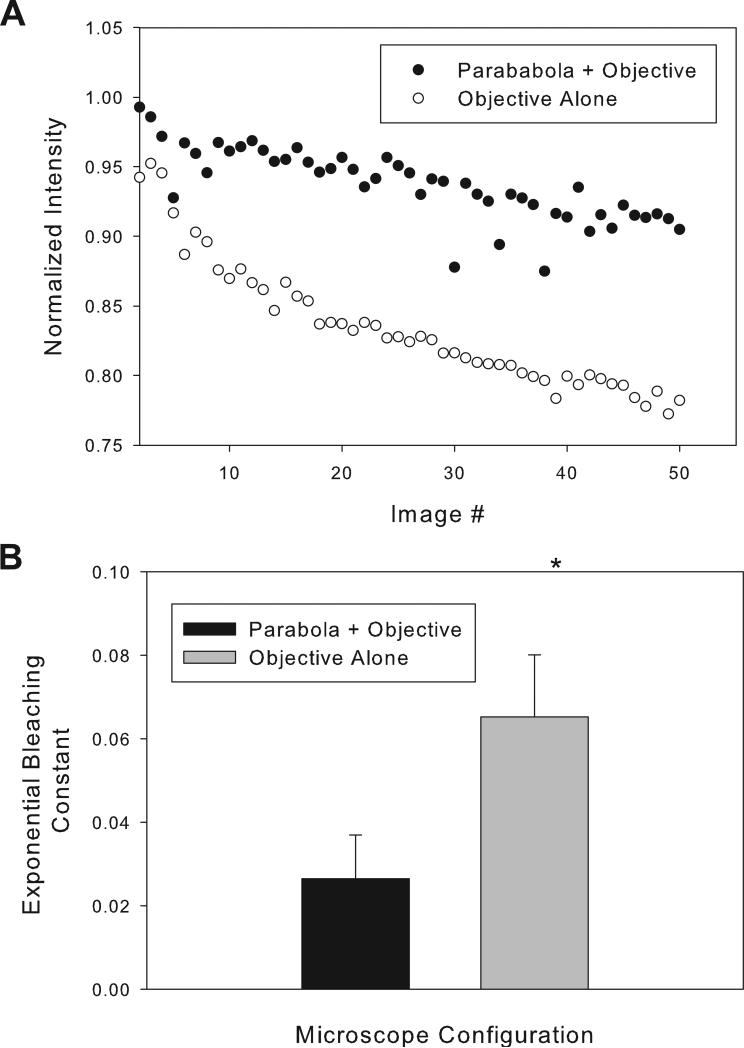
We describe a compact, non-contact design for a total emission detection (c-TED) system for intra-vital multiphoton imaging. To conform to a standard upright two-photon microscope design, this system uses a parabolic mirror surrounding a standard microscope objective in concert with an optical path that does not interfere with normal microscope operation. The non-contact design of this device allows for maximal light collection without disrupting the physiology of the specimen being examined. Tests were conducted on exposed tissues in live animals to examine the emission collection enhancement of the c-TED device compared to heavily optimized objective-based emission collection. The best light collection enhancement was seen from murine fat (5×-2× gains as a function of depth), whereas murine skeletal muscle and rat kidney showed gains of over two and just under twofold near the surface, respectively. Gains decreased with imaging depth (particularly in the kidney). Zebrafish imaging on a reflective substrate showed close to a twofold gain throughout the entire volume of an intact embryo (approximately 150 μm deep). Direct measurement of bleaching rates confirmed that the lower laser powers, enabled by greater light collection efficiency, yielded reduced photobleaching in vivo. The potential benefits of increased light collection in terms of speed of imaging and reduced photo-damage, as well as the applicability of this device to other multiphoton imaging methods is discussed.

Keywords: Imaging; light collection; two-photon microscopy.


Figures

Figure 1
Figure 1
The parabolic mirror assembly surrounds an objective on a regular commercially available upright two-photon microscope. Emission light collected by the parabola and the objective is reflected from a dichroic mirror in the turret and sent through an emission filter and focusing lens onto a wide-are PMT. The inset picture shows the relationship of the parabola to the sample for an experiment involving imaging of a leg muscle (Tibialis anterior). The dichroic mirror can be moved by a slider to enable standard wide-field imaging. Also shown is the thread system that allows focusing of the parabola.
Figure 2
Figure 2
Comparison of the compact TED collection efficiency and that of the objective alone for imaging fat and mixed tissue in murine skeletal muscle near the knee. A. Maximum intensity projection for the first 25 microns collected with the parabola and objective (left) and the objective alone (right). B. Single imaging plane from approximately 20 microns into the image stack depicted in A. Details of the imaging parameters can be found in “Materials and Methods”.
Figure 3
Figure 3
Raw data of pixel intensities and gain by depth for the image stack depicted in figure 2A. A and B represent pixel histograms of intensities for the entire image stack depicted in 2A for c-TED and for the objective alone microscope configurations, respectively. C. Gain in collection efficiency as a function of imaging depth for the fat-dominated, mixed tissue depicted image shown in figure 2A.
Figure 4
Figure 4
Comparison of the c-TED collection efficiency (left) with that of the objective alone (right) for imaging three different types of tissues. A. Maximum intensity projections for a depth of 50 μm into skeletal for blood vessels labeled with di-8-ANNEPPS. B. Maximum intensity projections through the entire thickness (approximately 150 μm) of a zebrafish embryo expressing GFP. C. Maximum intensity projections for the first 25 μm through a live rat kidney labeled with di-8-ANNEPPS in the vasculature. Details of the imaging parameters can be found in “Materials and Methods”.
Figure 5
Figure 5
The relationship between gain versus imaging depth. A. Gain versus depth for two mammalian tissues and a zebrafish embryo imaged in vivo. The zebrafish was imaged through its whole thickness (approximately 150 μm). The skeletal muscle was imaged to a depth of 300 μm whereas the kidney (a much more absorptive tissue) yielded useful images to a depth of approximately 200 μm. B. Linear false color montage images from various depths in a rat kidney for both the full c-TED and the objective alone configurations. The numbers in the top row of images correspond to the maximum intensity value in the cTED configuration and were used to byte-scale the images (to the false color inset on the right) for comparison between the two microscope configurations. Details of the analysis and the imaging parameters can be found in “Materials and Methods”.
Figure 6
Figure 6
Comparison of bleaching rates between the c-TED system and the objective alone for tetra methyl rhodamine (TMRM) labeled murine skeletal muscle imaged in vivo. Initial intensities were matched by changing excitation power. A. Raw intensity values over time for c-TED and the objective-alone microscope configurations from one representative experiment. B. Average calculated decay constants for repeated experiments (n=4). Details of the analysis and the imaging parameters can be found in “Materials and Methods”.

Similar articles

Cited by

References

    1. Bakalar M, Schroeder JL, Pursley R, Pohida TJ, Glancy B, Taylor J, Chess D, Kellman P, Xue H, Balaban RS. Three-dimensional motion tracking for high-resolution optical microscopy, in vivo. J Microsc. 2012;246:237–247. - PMC - PubMed
    1. Centonze VE, White JG. Multiphoton excitation provides optical sections from deeper within scattering specimens than confocal imaging. Biophys J. 1998;75:2015–2024. - PMC - PubMed
    1. Combs CA, Smirnov A, Chess D, McGavern DB, Schroeder JL, Riley J, Kang SS, Lugar-Hammer M, Gandjbakhche A, Knutson JR, Balaban RS. Optimizing multiphoton fluorescence microscopy light collection from living tissue by noncontact total emission detection (epiTED). J Microsc. 2011;241:153–161. - PMC - PubMed
    1. Combs CA, Smirnov AV, Riley JD, Gandjbakhche AH, Knutson JR, Balaban RS. Optimization of multiphoton excitation microscopy by total emission detection using a parabolic light reflector. J Microsc. 2007;228:330–337. - PubMed
    1. Crosignani V, Dvornikov A, Aguilar JS, Stringari C, Edwards R, Mantulin WW, Gratton E. Deep tissue fluorescence imaging and in vivo biological applications. J Biomed Opt. 2012;17:116023. - PMC - PubMed

Publication types

LinkOut - more resources