Review
. 2014;21(6):653-62.
doi: 10.2174/0929867320666131119151136.

Mechanisms of epileptiform synchronization in cortical neuronal networks

Affiliations
Review

Mechanisms of epileptiform synchronization in cortical neuronal networks

M Avoli. Curr Med Chem. 2014.

Abstract

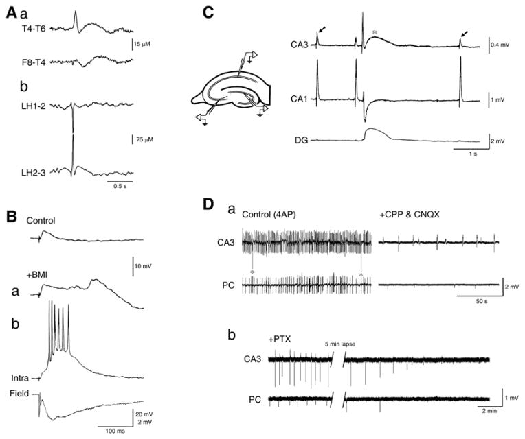
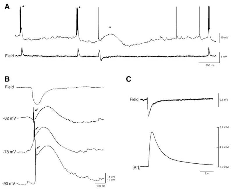
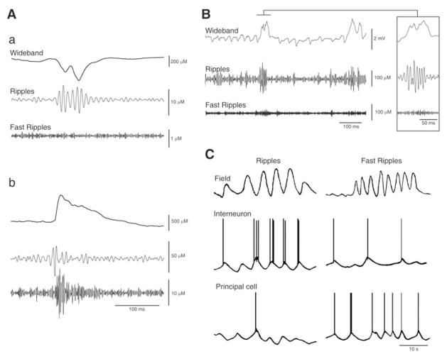
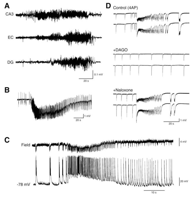
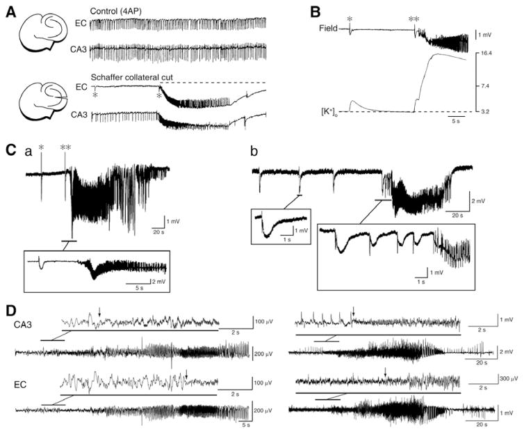
Neuronal synchronization supports different physiological states such as cognitive functions and sleep, and it is mirrored by identifiable EEG patterns ranging from gamma to delta oscillations. However, excessive neuronal synchronization is often the hallmark of epileptic activity in both generalized and partial epileptic disorders. Here, I will review the synchronizing mechanisms involved in generating epileptiform activity in the limbic system, which is closely involved in the pathophysiogenesis of temporal lobe epilepsy (TLE). TLE is often associated to a typical pattern of brain damage known as mesial temporal sclerosis, and it is one of the most refractory adult form of partial epilepsy. This epileptic disorder can be reproduced in animals by topical or systemic injection of pilocarpine or kainic acid, or by repetitive electrical stimulation; these procedures induce an initial status epilepticus and cause 1-4 weeks later a chronic condition of recurrent limbic seizures. Remarkably, a similar, seizure-free, latent period can be identified in TLE patients who suffered an initial insult in childhood and develop partial seizures in adolescence or early adulthood. Specifically, I will focus here on the neuronal mechanisms underlying three abnormal types of neuronal synchronization seen in both TLE patients and animal models mimicking this disorder: (i) interictal spikes; (ii) high frequency oscillations (80-500 Hz); and (iii) ictal (i.e., seizure) discharges. In addition, I will discuss the relationship between interictal spikes and ictal activity as well as recent evidence suggesting that specific seizure onsets in the pilocarpine model of TLE are characterized by distinctive patterns of spiking (also termed preictal) and high frequency oscillations.


Conflict of interest statement

CONFLICT OF INTEREST

The author(s) confirm that this article content has no conflicts of interest.

Figures

Fig. 1
Fig. 1
A: Electrographic features of interictal spikes recorded from a TLE patient with skull (a) and depth (b) electrodes; EEG recordings kindly provided by Drs. P. Perucca, F. Dubeau and J. Gotman at the Montreal Neurological Institute & Hospital. B: Changes of the responses generated by a neocortical neuron recorded intracellularly with a K-acetate-filled microelectrode in a human brain slice following single shock stimuli (triangle) under control conditions (Control) and during application of medium containing the GABAA receptor antagonist bicuculline methiodide (+BMI); trace in a was obtained shortly after the beginning of BMI application, and it shows the emergence of a late depolarizing potential that coincides with the time occupied under control conditions by a hyperpolarizing IPSP; traces shown in b, which were obtained after several minutes of BMI application, illustrate the interictal-like response recorded with a field electrode (Field), and the simultaneous intracellular paroxysmal depolarizing shift (Intra). C: Simultaneous field potential recordings obtained from an isolated hippocampal slice during application of 4-aminopyridine (4AP) show a pattern of “fast”, CA3-driven interictal spikes (arrows) that are not present in the dentate gyrus (DG) along with a “slow” interictal discharge (asterisk) that is recorded from all hippocampal regions. D: Simultaneous field potential recordings obtained from the CA3 subfield and the perirhinal cortex (PC) in a brain slice during application of 4AP (Control), after addition of glutamatergic receptor antagonists (CPP & CNQX), and after further addition of the GABAA receptor antagonist picrotoxin (PTX). Note that spontaneous field events continue to occur synchronously in the presence of glutamatergic receptor antagonists while the fast, CA3-driven interictal activity is abolished; note also that the glutamatergic-independent activity recorded from the CA3 subfield and the perirhinal cortex is abolished by picrotoxin.
Fig. 2
Fig. 2
A: Intracellular (K-acetate-filled microelectrode, Intra) and field potential (Field) recordings obtained from the CA3 subfield during 4AP application. Note that the intracellular counterpart of the “fast” interictal discharges recorded from a CA3 pyramidal cell consists of depolarizations that trigger bursts of fast action potentials while the counterpart of the “slow” interictal spike is characterized by a slow depolarization with a single fast action potential. B: Field and intracellular characteristics of the slow interictal discharge recorded from the CA3 subfield during application of 4AP and glutamatergic antagonists; note that depolarizing the resting membrane potential with steady current injection (−62 mV trace) discloses an initial hyperpolarizing component during which ectopic action potentials occur (arrow) as well as that similar ectopic “spikes” occur at more polarized membrane potentials (− 78 and −90 mV, arrows in both traces). Neurons recorded intracellularly in A and B were regularly firing, presumptive pyramidal cells. C: Simultaneous field potential and extracellular [K+] recordings of the glutamatergic independent interictal spike generated by entorhinal cortex neuronal networks during concomitant application of 4AP and glutamatergic antagonists; note that the extracellular [K+] increases up to approx. 5.0 mM from a resting concentration of 3.2 mM shortly after the negative peak of the field potential.
Fig. 3
Fig. 3
A: EEG recordings obtained with a depth electrode placed in the entorhinal cortex of two pilocarpine-treated epileptic rats show HFOs in the ripple (a) and fast ripple (b) band occurring in coincidence with interictal spikes. B: EEG recording from the CA3 subfield of a pilocarpine-treated epileptic rat approx. 6 s after seizure onset shows the occurrence of a ripple (also illustrated at high time resolution in the inset). In both A and B panels the raw EEG signal (Wideband trace) was bandpass filtered in the 80–200 Hz (Ripples trace) and in the 250–500 Hz (Fast Ripples trace) frequency range. C: Model figure summarizing the hypothetical intracellular activity of a principal cell and of an interneuron during a ripple and a fast ripple. Note that the principal cell generates hyperpolarizing (presumably GABAergic) potentials during the ripple while it fires action potentials during the fast ripple; in contrast, the inhibitory interneuron fires in phase during the ripple but it does so randomly during the fast ripple.
Fig. 4
Fig. 4
A: Seizure recorded with depth EEG electrodes placed in CA3, entorhinal cortex (EC) and dentate gyrus (DG) from a pilocarpine-treated epileptic rat. B: Electrographic seizure-like discharge recorded from the entorhinal cortex in a rat brain slice superfused with 4AP-containing medium. C: Simultaneous field (Field) and intracellular (−68 mV) recordings from the entorhinal cortex of a rat brain slice super-fused with Mg2+-free medium. The intracellular microelectrode was filled with KCl. D: Epileptiform activity recorded from two field recording electrodes placed in the rat cingulate cortex is modulated by μ-opioid receptors; note that the ictal activity induced by 4AP (Control) no longer persists after bath application of the μ-opioid agonist DAGO; this effect is reversed by the opioid antagonist naloxone.
Fig. 5
Fig. 5
A: Changes induced by Schaffer collateral cut on the epileptiform activity recorded from the CA3 and entorhinal cortex from a combined mouse brain slice. Under control conditions fast CA3-driven interictal activity is present in both areas; however, cutting the Schaffer collateral prevents CA3-driven interictal activity to propagate to the entorhinal cortex and uncovers ictal discharge (dotted line) along with slow interictal events (asterisks) in this area. B: Field and extracellular [K+] recordings during a slow interictal discharge and during the onset of a seizure-like discharge in the rat entorhinal cortex during 4AP application; note that the slow interictal spike (asterisk) is associated with an increase in extracellular [K+] that is smaller than what occurring in coincidence with the initial spike leading to ictal activity (double asterisk). Note also the much larger elevation in extracellular [K+] associated with the overt ictal discharge. C: Ictal discharges recorded from the entorhinal cortex in two brain slices that were superfused with 4AP containing medium. Note in a the occurrence of “slow” interictal discharges as well as that ictal onset is characterized by low-voltage oscillations at 10 Hz that follow by approx. 6 s a slow interictal event. In contrast, the onset of the ictal event shown in b is characterized by a pattern of acceleration of the “slow” interictal events. D: EEG recordings obtained from the hippocampus (CA3) and the entorhinal cortex (EC) of two pilocarpine-treated epileptic rats show onset patterns similar to those seen in vitro in C. These patterns consist of a spike followed by low-amplitude, high-frequency activity (asterisk in a) or of a series of spikes at approx. 1.5 Hz (asterisks in b) that are also followed by high frequency activity. Note that in both cases the high frequency activity (which signals the onset of the electrographic seizure) was observed first in the CA3 area.

Similar articles

Cited by

References

    1. Niedermeyer E, Lopes Da Silva FH. Electroencephalography: Basic Principles, Clinical Applications, and Related Fields. Lippincott Williams & Wilkins; 2005.
    1. Gloor P. The temporal lobe and limbic system. New York: Oxford University Press; 1997.
    1. Jefferys JGR, Jiruska P, de Curtis M, Avoli M. Limbic Network Synchronization and Temporal Lobe Epilepsy. In: Noebels JL, Avoli M, Rogawski MA, Olsen RW, Delgado-Escueta AV, editors. Jasper’s Basic Mechanisms of the Epilepsies [Internet] 4. Bethesda (MD): National Center for Biotechnology Information (US); 2012. - PubMed
    1. Engel J, Jr, McDermott MP, Wiebe S, Langfitt JT, Stern JM, Dewar S, Sperling MR, Gardiner I, Erba G, Fried I, Jacobs M, Vinters HV, Mintzer S, Kieburtz K Early Randomized Surgical Epilepsy Trial (ERSET) Study Group. Early surgical therapy for drug-resistant temporal lobe epilepsy: a randomized trial. JAMA. 2012;307:922–930. - PMC - PubMed
    1. Semah F, Picot MC, Adam C, Broglin D, Arzimanoglou A, Bazin B, Cavalcanti D, Baulac M. Is the underlying cause of epilepsy a major prognostic factor for recurrence? Neurology. 1998;51:1256–1262. - PubMed

Publication types