. 2014 Feb 1;189(3):314-24.
doi: 10.1164/rccm.201302-0302OC.

Regulation of hypoxia-induced pulmonary hypertension by vascular smooth muscle hypoxia-inducible factor-1α

Affiliations

Regulation of hypoxia-induced pulmonary hypertension by vascular smooth muscle hypoxia-inducible factor-1α

Molly K Ball et al. Am J Respir Crit Care Med. .

Abstract

Rationale: Chronic hypoxia induces pulmonary vascular remodeling, pulmonary hypertension, and right ventricular hypertrophy. At present, little is known about mechanisms driving these responses. Hypoxia-inducible factor-1α (HIF-1α) is a master regulator of transcription in hypoxic cells, up-regulating genes involved in energy metabolism, proliferation, and extracellular matrix reorganization. Systemic loss of a single HIF-1α allele has been shown to attenuate hypoxic pulmonary hypertension, but the cells contributing to this response have not been identified.

Objectives: We sought to determine the contribution of HIF-1α in smooth muscle on pulmonary vascular and right heart responses to chronic hypoxia.

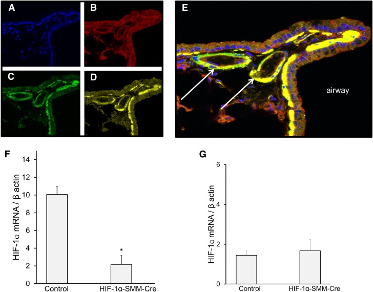
Methods: We used mice with homozygous conditional deletion of HIF-1α combined with tamoxifen-inducible smooth muscle-specific Cre recombinase expression. Mice received either tamoxifen or vehicle followed by exposure to either normoxia or chronic hypoxia (10% O2) for 30 days before measurement of cardiopulmonary responses.

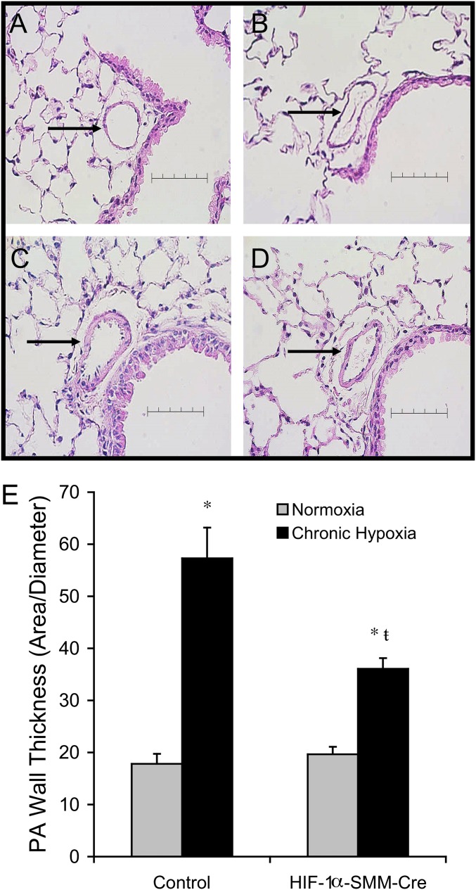
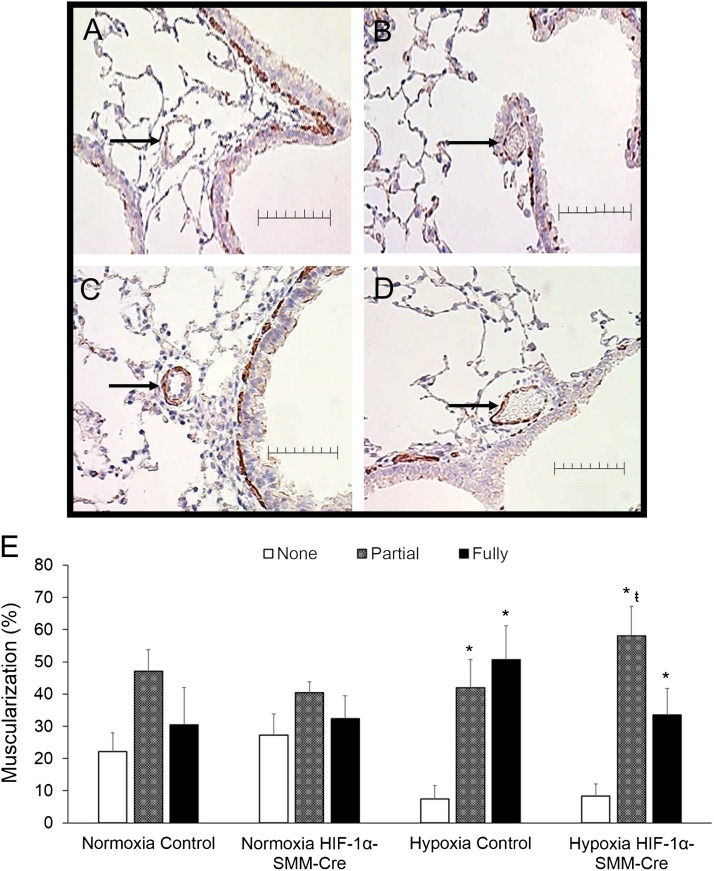
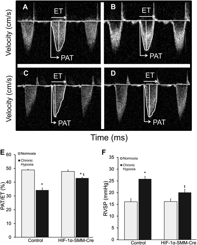
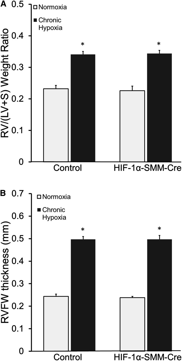
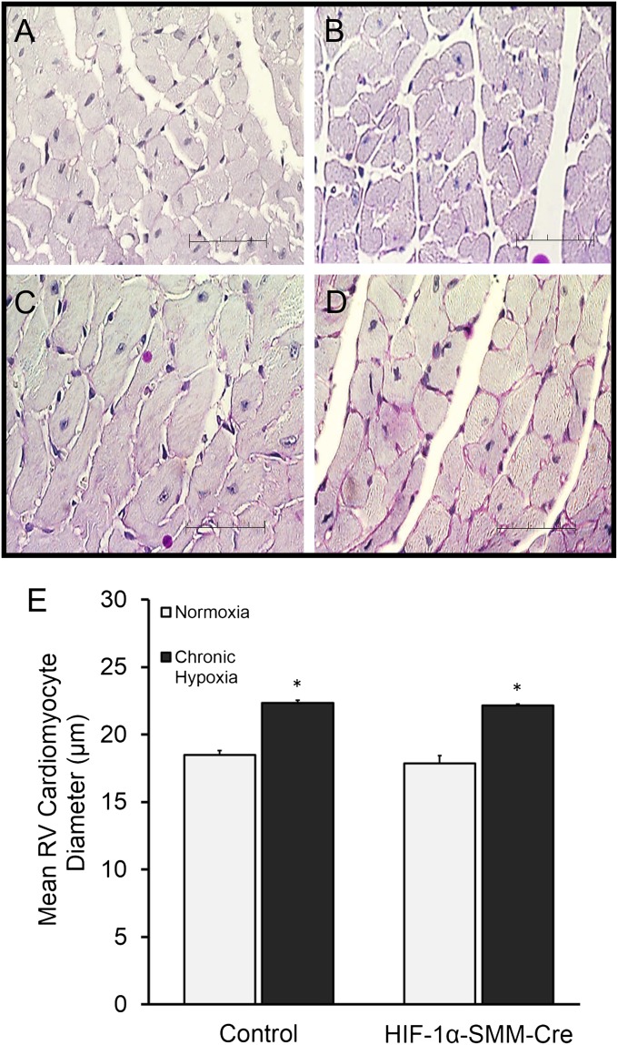
Measurements and main results: Tamoxifen-induced smooth muscle-specific deletion of HIF-1α attenuated pulmonary vascular remodeling and pulmonary hypertension in chronic hypoxia. However, right ventricular hypertrophy was unchanged despite attenuated pulmonary pressures.

Conclusions: These results indicate that HIF-1α in smooth muscle contributes to pulmonary vascular remodeling and pulmonary hypertension in chronic hypoxia. However, loss of HIF-1 function in smooth muscle does not affect hypoxic cardiac remodeling, suggesting that the cardiac hypertrophy response is not directly coupled to the increase in pulmonary artery pressure.


Figures

Figure 1.
Figure 1.
Smooth muscle–specific hypoxia-inducible factor (HIF)-1α deletion. Tamoxifen induction of Cre recombinase activity in smooth muscle; confocal microscopy images of mouse lung tissue slices at ×40 magnification. (A) DAPI counterstain. (B) Labeled mTmG-reporter constitutively expressing red fluorescence in all cell types. (C) Tamoxifen treatment causes expression to change from red to green fluorescence in cells expressing Cre. (D) Immunofluorescence labeling of smooth muscle actin (yellow) in pulmonary vasculature and airways. (E) Colocalization of yellow and green fluorescence, confirming activation of Cre recombinase in smooth muscle. Arrows identify small pulmonary arteries adjacent to a labeled airway. (F) HIF-1α mRNA message by reverse-transcriptase polymerase chain reaction in normoxic smooth muscle cells after in vivo tamoxifen treatment, confirming effective tissue-specific knock-out. n = 2–3/group; *P < 0.05 versus control. (G) HIF-1α mRNA message by reverse-transcriptase polymerase chain reaction in normoxic right ventricular tissue after in vivo tamoxifen treatment, demonstrating that HIF-1α mRNA expression is retained in cardiomyocytes after HIF-1α deletion in smooth muscle.
Figure 2.
Figure 2.
Vascular remodeling in small pulmonary arteries after chronic hypoxia exposure. Representative hematoxylin and eosin–stained mouse lung tissue sections. Arrows identify a small pulmonary artery adjacent to a labeled airway. Scale bar = 80 μm. (A) Normoxic control. (B) Normoxic hypoxia-inducible factor (HIF)-1α–Cre recombinase under the control of a smooth muscle–specific promoter (SMM-Cre). (C) Chronic hypoxia. (D) Chronic hypoxia HIF-1α–SMM-Cre. (E) Quantification of pulmonary vascular remodeling in chronic hypoxia by wall thickness demonstrates attenuated remodeling in HIF-1α–SMM-Cre mice. P < 0.05 versus normoxic control (*) or versus hypoxic control (ŧ). PA = pulmonary artery.
Figure 3.
Figure 3.
Muscularization in distal pulmonary arteries after chronic hypoxia exposure. (A–D) Representative α-smooth muscle actin–stained mouse lung tissue. Arrows identify a pulmonary artery with diameter less than 50 μm. Scale bar = 50 μm. (A) normoxia control. (B) Normoxia hypoxia-inducible factor (HIF)-1α–Cre recombinase under the control of a smooth muscle–specific promoter (SMM-Cre). (C) Chronic hypoxia. (D) Chronic hypoxia HIF-1α–SMM-Cre. (E) Hypoxia-induced muscularization of distal pulmonary arteries (<50-μm diameter) by characterization as nonmuscularized, partially muscularized, and fully muscularized. Chronic hypoxia induced distal muscularization in both groups; however, less full muscularization developed in HIF-1α–SMM-Cre mice relative to chronic hypoxic controls, indicating fewer vessels with complete circumferential muscularization. P < 0.05 versus corresponding group nonmuscularized vessels (*) or versus corresponding group fully muscularized vessels (ŧ).
Figure 4.
Figure 4.
Pulmonary hypertension after exposure to chronic hypoxia. (A–D) Pulse-wave Doppler images across the pulmonary outflow tract: (A) normoxic control, (B) normoxic hypoxia-inducible factor (HIF)-1α–Cre recombinase under the control of a smooth muscle–specific promoter (SMM-Cre), (C) chronic hypoxia, (D) chronic hypoxia HIF-1α–SMM-Cre. The time to peak flow acceleration across the pulmonary valve decreases with increasing pulmonary artery pressures; the ratio of pulmonary acceleration time to total pulmonary ejection time (PAT/ET) varies inversely with pulmonary artery pressure. (E) Pulmonary hypertension after exposure to chronic hypoxia. PAT/ET demonstrates pulmonary hypertension after chronic hypoxia, which is attenuated in HIF-1α–SMM-Cre mice. (F) Right ventricular systolic pressure (RVSP) after exposure to chronic hypoxia. P < 0.05 versus normoxic control (*) or versus hypoxic control (ŧ).
Figure 5.
Figure 5.
Cardiac remodeling in chronic hypoxia. (A) Fulton index (RV/[LV+S]) measurement of right ventricular mass. Mice exposed to chronic hypoxia develop increased right heart mass compared with normoxic control littermates. (B) Echocardiographic measurement of right ventricular free wall (RVFW) thickness additionally demonstrates development of right ventricular hypertrophy in all mice after chronic hypoxia. *P < 0.05 versus normoxic control. HIF = hypoxia-inducible factor; LV = left ventricle; RV = right ventricle; S = interventricular septum; SMM-Cre = Cre recombinase under the control of a smooth muscle–specific promoter.
Figure 6.
Figure 6.
Hypertrophy of right ventricular cardiomyocytes in chronic hypoxia. Representative periodic acid Schiff–stained mouse right ventricular tissue sections. (A) Normoxia control. (B) Normoxia hypoxia-inducible factor (HIF)-1α–Cre recombinase under the control of a smooth muscle–specific promoter (SMM-Cre). (C) Hypoxia control. (D) Hypoxia HIF-1α–SMM-Cre. Scale bar = 50 μm. (E) Mice exposed to chronic hypoxia exhibit increased right ventricular fiber diameter compared with normoxic control littermates. Scale bar = 50 μm. RV = right ventricle. *P < 0.05 versus normoxic control.

Comment in

References

    1. Strange C, Highland KB. Pulmonary hypertension in interstitial lung disease. Curr Opin Pulm Med. 2005;11:452–455. - PubMed
    1. Roy R, Couriel JM. Secondary pulmonary hypertension. Paediatr Respir Rev. 2006;7:36–44. - PubMed
    1. Stenmark KR, Abman SH. Lung vascular development: implications for the pathogenesis of bronchopulmonary dysplasia. Annu Rev Physiol. 2005;67:623–661. - PubMed
    1. Naeije R. Pulmonary hypertension and right heart failure in chronic obstructive pulmonary disease. Proc Am Thorac Soc. 2005;2:20–22. - PubMed
    1. Stern RC, Borkat G, Hirschfeld SS, Boat TF, Matthews LW, Liebman J, Doershuk CF. Heart failure in cystic fibrosis. Treatment and prognosis of cor pulmonale with failure of the right side of the heart. Am J Dis Child. 1980;134:267–272. - PubMed

Publication types

MeSH terms

Substances