. 2014 Apr;20(7-8):1271-84.
doi: 10.1089/ten.TEA.2012.0773.

Differentiation of human adipose-derived stem cells into neuron-like cells which are compatible with photocurable three-dimensional scaffolds

Affiliations

Differentiation of human adipose-derived stem cells into neuron-like cells which are compatible with photocurable three-dimensional scaffolds

Shane Gao et al. Tissue Eng Part A. 2014 Apr.

Abstract

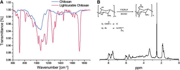
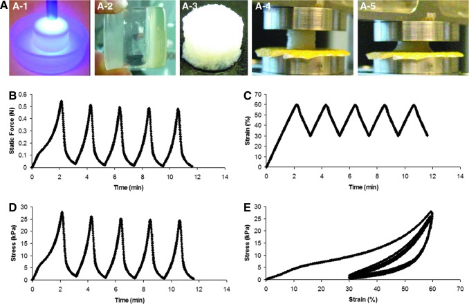
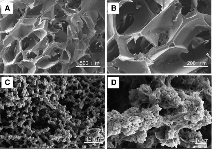
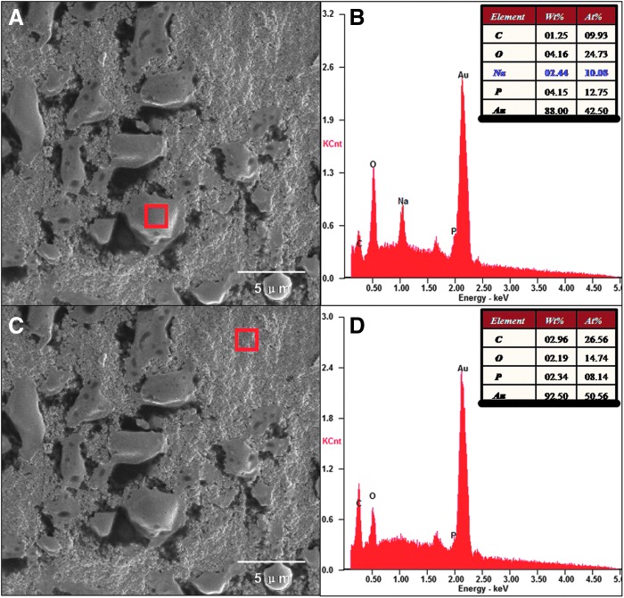
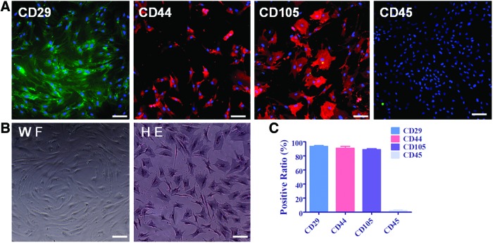
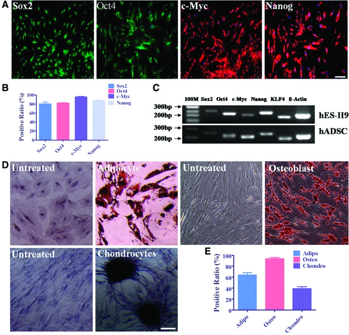
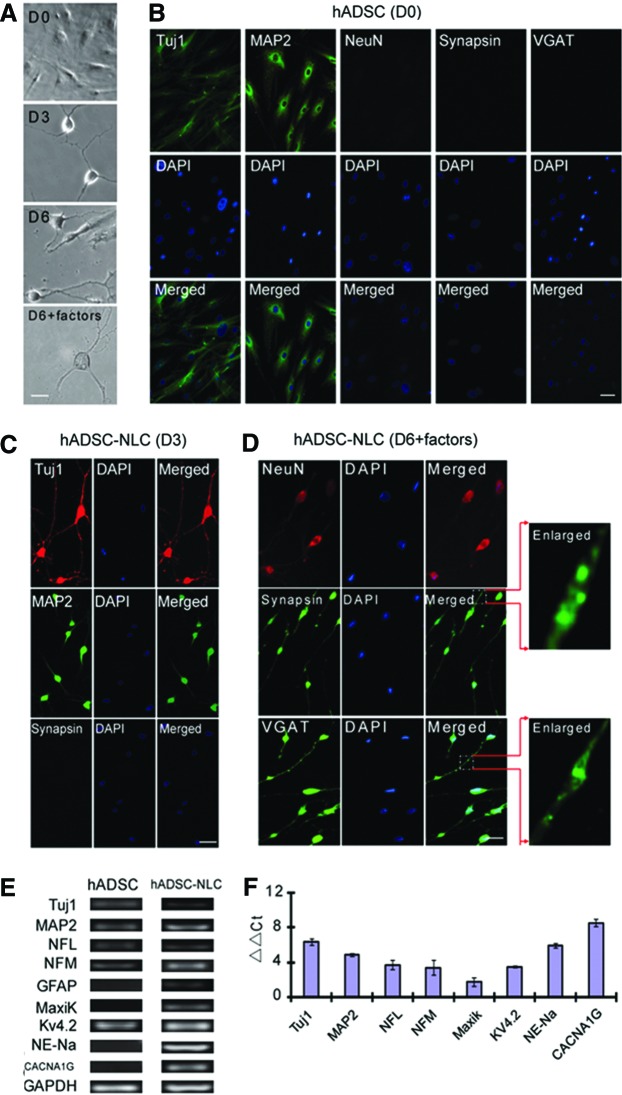
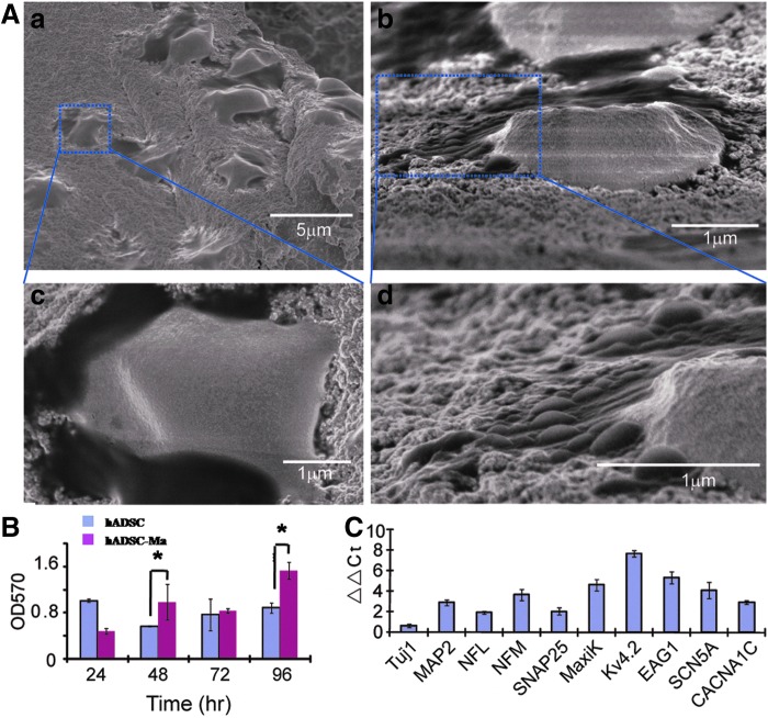
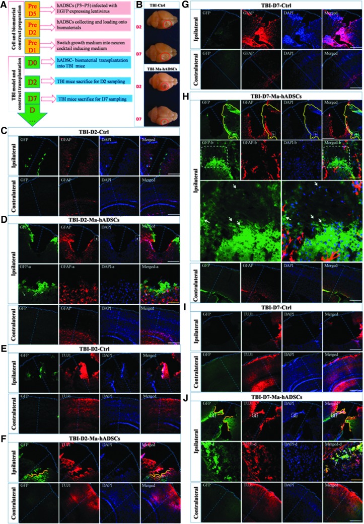
Multipotent human adipose-derived stromal/stem cells (hADSCs) hold a great promise for cell-based therapy for many devastating human diseases, such as spinal cord injury and stroke. If exogenous hADSCs can be cultured in a three-dimensional (3D) scaffold with effective proliferation and differentiation capacity, it will better mimic the in vivo environment, which will have profound impact on the therapeutic application of hADSCs. In this study, a group of elastic-dominant, porous bioscaffolds from photocurable chitosan and gelatin were fabricated and proven to be biocompatible with both hADSCs and hADSC-derived neuron-like cells (hADSC-NLCs) in vitro. The identity of harvested hADSCs was confirmed by their positive immunostaining of mesenchymal stem cell surface markers, CD29, CD44, and CD105, and also positive expression of stem markers, Sox-2, Oct-4, c-Myc, Nanog, and Klf4. Their multipotency was further confirmed by trilineage differentiation of hADSCs toward adipocyte, osteoblast, and chondrocyte. It was found that hADSCs could be conditioned to differentiate into neurons in vitro as determined by immunostaining the markers of Tuj1, MAP2, NeuN, and Synapsin. The hADSCs and hADSC-NLCs were proven to be biocompatible with 3D scaffold, which actually facilitated the proliferation and differentiation of hADSCs in vitro, by MTT assay and their neuronal gene expression profiling. Moreover, hADSC-NLCs, which were mixed with 3D scaffold and transplanted into traumatic brain injury mouse model, survived in vivo and led to the better repair of the damaged brain area. The immunohistochemical studies revealed that 3D scaffold indeed improved the viability of transplanted cells, their ability to incorporate into the in vivo neural circuit, and their capacity for tissue repair. This study indicates that hADSCs would have great therapeutic application potential as seeding cells for in vivo transplantation to treat various neurological diseases when co-applied with porous chitosan/gelatin bioscaffolds.


Figures

<b>FIG. 1.</b>
FIG. 1.
FTIR-ATR (A) and 1H NMR (B) characterization of light-curable chitosan.
<b>FIG. 2.</b>
FIG. 2.
(A) Three-dimensional (3D) scaffolds fabrication scheme (A-1, A-2, and A-3: UV exposure; A-4 and A-5: compression test) and cyclic compression test on the chitosan–gelatin hybrid scaffolds (5%–5%) at a constant strain rate of 1 mm/min and a strain range of 30%–60%. (B) Static force versus time; (C) strain versus time; (D) stress versus time; and (E) stress–strain curve.
<b>FIG. 3.</b>
FIG. 3.
Scanning electron microscopy (SEM) morphology of the 3D scaffolds (5% gelatin-5% light-curable chitosan) made with NaCl (250–380 μm) as porgen: (A, B) top view; (C, D) cross section with nanostructures, such as gelatin beads.
<b>FIG. 4.</b>
FIG. 4.
SEM images of the 3D scaffolds coupled with day 6 human adipose-derived stromal/stem cell (hADSC)-neuron-like cells (NLCs) (A, C). The corresponding energy dispersive X-ray analyzer (EDS) spectrum on cell surface (A) and scaffold surface (C) showed energetic lines for carbon, oxygen, Na+ and phosphrous (B, D).
<b>FIG. 5.</b>
FIG. 5.
Characterization of hADSCs by cell morphology and CD surface markers. hADSCs grow swirling with spindle-like morphology. (A) Shows more than 85% of hADSCs at P3 positively express CD29, CD44, and CD105 and less than 5% of hADSCs express CD45. P3 of hADSCs plated on plastic culture dish in Dulbecco's modified Eagle's medium (DMEM)/F12 medium supplemented with 10% fetal bovine serum (FBS) taken under white field (WF) and hematoxylin and eosin (H&E) staining to observe hADSC morphology and nuclear was shown on (B). (C) Shows the quantification of CD-positive ratio of cells by taking average of at least five microscope fields. Scale bar=100 μm. Color images available online at www.liebertpub.com/tea
<b>FIG. 6.</b>
FIG. 6.
Stem cell transcriptional factor expression determination and trilineage differentiation of hADSCs. Immunocytochemistry staining with Sox2, Oct4, c-Myc, and Nanog showed that more than 80% of hADSCs positively express these markers, which is consistent with their mRNA level expression comparing with human ES cell line of H9 (A–C). hADSCs can be specifically induced into adipocytes stained with Oil Red O, osteoblast cells stained with Alizarin, and chondrocytes stained with Toluidine blue by culturing in certain and specific differentiating medium for 14 days (D). The untreated means hADSCs were cultured in normal medium and performed later staining in parallel with those cultured in the differentiating medium. The medium was changed every 3 days. The differentiation positive ratio of trilineages was collected by taking average of at least five microscope view fields (E). Scale bar=100 μm. Color images available online at www.liebertpub.com/tea
<b>FIG. 7.</b>
FIG. 7.
hADSCs were induced into (A) NLCs with morphology changing continuously as time lapse from day 0 (D0) to day 6 (D6). (B) Without any treatment, hADSCs positively express Tuj1 and MAP2 and negatively express NeuN, Synapsin1/2, and vGAT. After 3-day incubation with cocktail medium, most hADSCs-NLCs positively expressed Tuj1, MAP2 but not Synapsin1/2 (C). After additional 3-day incubation with the cocktail medium supplemented with neurotrophic factors, most cells began to express neuron function-related markers of NeuN, Synapsin, and vGAT (D). qRT-PCR data demonstrated that neuron markers such as NFL, NFM, as well as function- and ion channel-related genes such as Kv4.2, NE-Na, and CACNA1G were all upregulated (E, F). Scale bar=25 μm for (A), scale bar=50 μm for (B–D). Color images available online at www.liebertpub.com/tea
<b>FIG. 8.</b>
FIG. 8.
hADSCs cultured on the 3D scaffold were subjected to the neuron cocktail medium with similar operation on the plastic dishes. After 6 day differentiation, hADSC-NLCs were detected the morphology by SEM (A). To monitor hADSC proliferation capability on this 3D scaffold material, MTT assay were carried out within 96 h at a time course of 24 h (B). Both hADSC-NLCs and hADSCs on 3D scaffold at D6 were subjected to trizol digestion for total RNA extraction. qRT-PCR were carried out to detect the neuron-related gene expression variance (C). ADSC-Ma means hADSCs cultured on the 3D scaffold material to compare with the hADSCs cultured on the plastic dishes. Two-way ANOVA with Bonferroni post-tests were performed for the data in (B) using GraphPad Prism version 5.00 for Windows, GraphPad Software (San Diego, CA), www.graphpad.com. *p<0.05.
<b>FIG. 9.</b>
FIG. 9.
Traumatic brain injury (TBI) mouse model for tracing the hADSC-NLC/chitosan/gelatin scaffold meanwhile using phosphate-buffered saline (PBS) as the control group (n=6). (A) Shows the protocols to perform this experiment; (B) shows the representative TBI brain morphology at different time points (D2 and D7) in each group (n=6). Brain dissection and histoimmunostaining with GFAP and TUJ1 were performed at time course of 2 days (D2) and 7 days (D7) after TBI surgery and cell material construct transplantation. In figures (C, E, G, I), TBI-D2-Ctrl and TBI-D7-Ctrl means TBI mice without treatment but PBS placebo injection as the control group sacrificed on D2 or D7 after TBI surgery. In figures (D, F, H, J), TBI-D2-Ma-hADSCs and TBI-D7-Ma-hADSCs means TBI mice transplanted with hADSC-NLC/chitosan/gelatin scaffold sacrificed on D2 or D7 after TBI surgery. Scale bar (yellow line=100 μm, white line=500 μm).

References

    1. Zuk P.A, Zuh M., Mizuno H., Huang J., Futrell J.W., Katz A.J., et al. . Multilineage cells from human adipose tissue: implications for cell-based therapies. Tissue Eng 7,211, 2001 - PubMed
    1. Gronthos S., Franklin D.M., Leddy H.A., Robey P.G., Storms R.W., and Gimble J.M.Surface protein characterization of human adipose tissue-derived stromal cells. J Cell Physiol 189,54, 2001 - PubMed
    1. Zuk P.A., Zhu M., Ashjian P., De Ugarte D.A., Huang J.I., Mizuno H., et al. . Human adipose tissue is a source of multipotent stem cells. Mol Biol Cell 13,4279, 2002 - PMC - PubMed
    1. Zhu Y., Liu T., Song K., Fan X., Ma X., and Cui Z.Adipose-derived stem cell: a better stem cell than BMSC. Cell Biochem Funct 26,664, 2008 - PubMed
    1. Denker A.E., Nicoll S.B., and Tuan R.S.Formation of cartilage-like spheroids by micromass cultures of murine C3H10T1/2 cells upon treatment with transforming growth factor beta-1. Differentiation 59,25, 1995 - PubMed

Publication types

MeSH terms