. 2014 Apr;129(1):120-9.
doi: 10.1111/jnc.12611. Epub 2013 Dec 6.

Ischemic neurons activate astrocytes to disrupt endothelial barrier via increasing VEGF expression

Affiliations

Ischemic neurons activate astrocytes to disrupt endothelial barrier via increasing VEGF expression

Ying-Na Li et al. J Neurochem. 2014 Apr.

Abstract

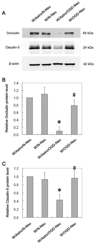
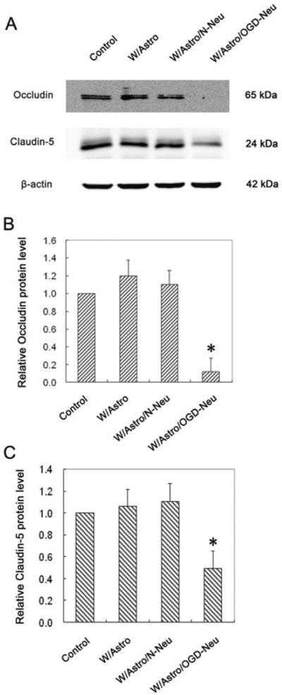
Blood-brain barrier (BBB) disruption occurring within the first few hours of ischemic stroke onset is closely associated with hemorrhagic transformation following thrombolytic therapy. However, the mechanism of this acute BBB disruption remains unclear. In the neurovascular unit, neurons do not have direct contact with the endothelial barrier; however, they are highly sensitive and vulnerable to ischemic injury, and may act as the initiator for disrupting BBB when cerebral ischemia occurs. Herein, we employed oxygen-glucose deprivation (OGD) and an in vitro BBB system consisting of brain microvascular cells and astrocytes to test this hypothesis. Neurons (CATH.a cells) were exposed to OGD for 3-h before co-culturing with endothelial monolayer (bEnd 3 cells), or endothelial cells plus astrocytes (C8-D1A cells). Incubation of OGD-treated neurons with endothelial monolayer alone did not increase endothelial permeability. However, when astrocytes were present, the endothelial permeability was significantly increased, which was accompanied by loss of occludin and claudin-5 proteins as well as increased vascular endothelial growth factor (VEGF) secretion into the conditioned medium. Importantly, all these changes were abolished when VEGF was knocked down in astrocytes by siRNA. Our findings suggest that ischemic neurons activate astrocytes to increase VEGF production, which in turn induces endothelial barrier disruption.

Keywords: VEGF; astrocyte; blood-brain barrier; neuron; oxygen-glucose deprivation.


Figures

Fig. 1
Fig. 1
OGD-treated neurons interact with astrocytes to increase endothelial monolayer permeability. Neuronal cells (CATH.a cells) grown on inserts were exposed to OGD for 3 hours and then co-cultured with astrocytes (C8-D1A cells) for 24 hours. After that, the neurons were removed and the astrocytes were incubated with endothelial monolayer (bEnd.3 cells) for another 24 hours. Endothelial monolayer permeability was assessed by quantitating the clearance of FITC-BSA from luminal compartment to abluminal compartment. The co-culture of OGD-treated neurons with astrocytes significantly increased endothelial barrier permeability. Data are expressed as mean ± SD, n = 3. *P < 0.05 vs Control. Control: endothelial monolayer alone; W/Astro: co-culturing endothelial monolayer with astrocytes; W/Astro/N-Neu: astrocytes were first co-cultured with normal neurons and then with endothelial monolayer; W/Astro/OGD-Neu: astrocytes were first co-cultured with OGD-treated neurons and then with endothelial monolayer
Fig. 2
Fig. 2
OGD-treated neurons interact with astrocytes to reduce occludin and claudin-5 protein levels in endothelial monolayer. Neuronal cells (CATH.a) grown on inserts were exposed to OGD for 3 hours and then co-cultured with astrocytes (C8-D1A) for 24 hours. After that, the neurons were removed and the astrocytes were incubated with endothelial monolayer (bEnd.3 cells) for another 24 hours before assessing occludin and claudin-5 protein levels by western blot. (A) Representative blots for occludin, claudin-5 and β-actin. The relative quantity of occludin protein (B) or claudin-5 protein (C) was calculated after normalization to β-actin. Co-culturing OGD-treated neurons with astrocytes significantly decreased occludin and claudin-5 protein levels in endothelial cells. Data represent the mean ± S.D, n = 3. *P < 0.05 vs Control. Control: endothelial monolayer alone; W/Astro: co-culturing endothelial monolayer with astrocytes; W/Astro/N-Neu: astrocytes were first co-cultured with normal neurons and then with endothelial monolayer; W/Astro/OGD-Neu: astrocytes were first co-cultured with OGD-treated neurons and then with endothelial monolayer
Fig. 3
Fig. 3
Astrocytes are required for ischemic neurons to exert its damaging effect on endothelial barrier. Neuronal cells (CATH.a cells) grown on inserts were exposed to OGD for 3 hours and then cultured for another 24 hours with or without astrocytes (C8-D1A cells). After that, the neurons were removed, and the astrocytes were incubated with endothelial monolayer for another 24 hours before assessing endothelial monolayer permeability to FITC-BSA. Data are expressed as mean ± SD, n = 3. *P < 0.05 vs W/Astro/N-Neu, #P < 0.05 vs W/Astro/OGD-Neu. W/Astro/N-Neu: astrocytes were first incubated with normal neurons and then with endothelial monolayer; W/N-Neu: Conditioned media collected from normal neurons and then with endothelial monolayer; W/Astro/OGD-Neu: astrocytes were first incubated with OGD-treated neurons and then with endothelial monolayer; W/OGD-Neu: incubating endothelial monolayer with the conditioned media collected from OGD-treated neurons
Fig. 4
Fig. 4
Removal of astrocytes abolishes the impact of OGD-treated neurons on occludin and claudin-5 protein levels in endothelial cells. Neuronal cells (CATH.a cells) grown on inserts were exposed or not to OGD for 3 hours and then co-cultured with astrocytes (C8-D1A cells) /or not for 24 hours. After that, the neurons were replaced by endothelial monolayer and incubated for another 24 hours before assessing occludin and claudin-5 protein levels by western blot. (A) Representative blots for occludin, claudin-5 and β-actin. The relative quantity of occludin protein (B) or claudin-5 protein (C) was calculated after normalization to β-actin. Data are expressed as mean ± SD, n = 3. *P < 0.05 vs W/Astro/N-Neu, #P < 0.05 vs W/Astro/OGD-Neu. W/Astro/N-Neu: astrocytes were first co-cultured with normal neurons and then with endothelial monolayer; W/N-Neu: incubating with normal neuron-conditioned media; W/Astro/OGD-Neu: astrocytes were first co-cultured with OGD-treated neurons and then with endothelial monolayer; W/OGD-Neu: incubating with conditioned media collected from OGD-treated neurons
Fig. 5
Fig. 5
OGD-treated neurons interact with astrocytes to increase VEGF production and secretion in astrocytes. The neuronal cells (CATH.a) were exposed or not to OGD for 3 hours before co-culturing with astrocytes (C8-D1A) for 24 hours. After co-culture, VEGF production and secretion were assessed by measuring its content in astrocytic extracts and conditioned media using western blot and ELISA, respectively. (A) OGD-treated neurons (OGD-Neuron), but not normal neurons (N-Neuron) induced a slight but significant increase in VEGF protein in astrocytic extract. Upper panel: representative blots for VEGF and β-actin. Lower panel: The relative quantity of VEGF protein was calculated after normalization to β-actin. Data are expressed as mean ± SD, n = 3, *P <0.05 vs Astrocyte (astrocytic culture alone). (B) VEGF secreted into the conditioned media was assessed using an ELISA kit. Data are expressed as mean ± SD, n = 3, *P <0.05 vs Astrocyte (astrocytic culture alone)
Fig. 6
Fig. 6
VEGF induces the disruption of endothelial barrier and the loss of occludin and claudin-5 proteins. Different concentrations of VEGF were added to the culture medium of the endothelial monolayer (bEnd.3 cells) with full confluence. After 24 hours incubation, the endothelial barrier permeability was assayed as described in “materials and methods” (A), and the protein levels of occludin and claudin-5 were assayed by western blot (B): the upper panel shows representative blots for occludin, claudin-5 and β-actin; the lower panel shows the relative quantity of occludin and claudin-5 protein after normalization to β-actin. *P < 0.05 vs Control (0 ng/mL group), n = 3.
Fig. 7
Fig. 7
VEGF mediates induced by co-culturing OGD-treated neurons with astrocytes. Astrocytes (C8-D1A) were transfected with control or VEGF siRNA for 72 hours before co-culturing them with OGD-treated neurons (CATH.a) and endothelial monolayer. (A) Transfection of astrcocytes with VEGF siRNA for 72 hours significantly reduced (~80% reduction) VEGF protein levels. The upper panel shows representative blots for VEGF and β-actin; the lower panel shows the relative quantity of VEGF protein after normalization to β-actin. *P < 0.05 vs Control siRNA. Knockdown of VEGF in astrocytes significantly blocked the impact of OGD-treated neurons on astrocytes and endothelial cells, as reflected by the findings that VEGF secretion was significantly reduced in astrocytes (B), occludin and claudin-5 protein loss was reversed in endothelial monolayer (C), and endothelial barrier permeability increase was significantly reduced (D). Controls are astrocytes alone (B), endothelial monolayer alone (C and D), respectively. Data are expressed as mean ± SD, *P < 0.05 vs Control, #P < 0.05 vs Control siRNA (ConsiRNA), n = 3.

Similar articles

Cited by

References

    1. Argaw AT, Asp L, Zhang J, et al. Astrocyte-derived VEGF-A drives blood-brain barrier disruption in CNS inflammatory disease. J Clin Invest. 2012;122:2454–2468. - PMC - PubMed
    1. Argaw AT, Gurfein BT, Zhang Y, Zameer A, John GR. VEGF-mediated disruption of endothelial CLN-5 promotes blood-brain barrier breakdown. Proc Natl Acad Sci U S A. 2009;106:1977–1982. - PMC - PubMed
    1. Armulik A, Genove G, Mae M, et al. Pericytes regulate the blood-brain barrier. Nature. 2010;468:557–561. - PubMed
    1. Ballabh P, Braun A, Nedergaard M. The blood-brain barrier: an overview: structure, regulation, and clinical implications. Neurobiol Dis. 2004;16:1–13. - PubMed
    1. Bickler PE, Donohoe PH. Adaptive responses of vertebrate neurons to hypoxia. J Exp Biol. 2002;205:3579–3586. - PubMed

Publication types

LinkOut - more resources