. 2014 Jan 1;189(1):16-29.
doi: 10.1164/rccm.201305-0874OC.

Regulation of IL-4 receptor signaling by STUB1 in lung inflammation

Affiliations

Regulation of IL-4 receptor signaling by STUB1 in lung inflammation

Qin Wei et al. Am J Respir Crit Care Med. .

Abstract

Rationale: IL-4Rα, the common receptor component for IL-4 and IL-13, plays a critical role in IL-4- and IL-13-mediated signaling pathways that regulate airway inflammation and remodeling. However, the regulatory mechanisms underlying IL-4Rα turnover and its signal termination remain elusive.

Objectives: To evaluate the role of STUB1 (STIP1 homology and U-Box containing protein 1) in regulating IL-4R signaling in airway inflammation.

Methods: The roles of STUB1 in IL-4Rα degradation and its signaling were investigated by immunoblot, immunoprecipitation, and flow cytometry. The involvement of STUB1 in airway inflammation was determined in vivo by measuring lung inflammatory cells infiltration, mucus production, serum lgE levels, and alveolar macrophage M2 activation in STUB1(-/-) mice. STUB1 expression was evaluated in airway epithelium of patients with asthma and lung tissues of subjects with chronic obstructive pulmonary disease.

Measurements and main results: STUB1 interacted with IL-4Rα and targeted it for ubiquitination-mediated proteasomal degradation, terminating IL-4 or IL-13 signaling. STUB1 knockout cells showed increased levels of IL-4Rα and sustained STAT6 activation, whereas STUB1 overexpression reduced IL-4Rα levels. Mice deficient in STUB1 had spontaneous airway inflammation, alternative M2 activation of alveolar macrophage, and increased serum IgE. STUB1 levels were increased in airways of subjects with asthma or chronic obstructive pulmonary disease, suggesting that up-regulation of STUB1 might be an important feedback mechanism to dampen IL-4R signaling in airway inflammation.

Conclusions: Our study identified a previously uncharacterized role for STUB1 in regulating IL-4R signaling, which might provide a new strategy for attenuating airway inflammation.


Figures

Figure 1.
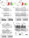
Figure 1.
STIP1 homology and U-Box–containing protein 1 (STUB1) down-regulates IL-4Rα levels. Expression of IL-4Rα was analyzed by immunoblot (A and C) or flow cytometry (B) in mouse embryonic fibroblasts (MEF), lung tissues (six mice per group), or tracheal epithelial cells from wild-type (WT), STUB1+/−, or STUB1−/− mice. (D) Immunoblot analysis of IL-4Rα in MEF infected with a recombinant lentivirus expressing STUB1 or a control lentivirus for 48 hours or in A549 cells cotransfected with plasmid encoding IL-4Rα together with either a control vector or plasmid encoding His-STUB1 for 48 hours. (E) A549 cells cotransfected with plasmids encoding Flag-IL-4Rα and STUB1 were pulsed with [35S]methionine-cysteine and chased with unlabeled medium at indicated time points. IL-4Rα was immunoprecipitated with α-Flag antibody, eluted, and analyzed by sodium dodecyl sulfate–polyacrylamide gel electrophoresis. Bands representing 35S-labeled IL-4Rα were quantified. (F) WT and STUB1−/− MEF were treated with 25 μM cyclohexamide for indicated periods and cell lysates were subjected to immunoblot to evaluate IL-4Rα. *Nonspecific band. Graph data are mean ± SD, *P < 0.05, **P < 0.01. Representative blots are shown from at least three independent experiments. GAPDH = glyceraldehyde phosphate dehydrogenase.
Figure 2.
Figure 2.
STIP1 homology and U-Box containing protein 1 (STUB1) interacts with IL-4Rα. (A) Cell lysates of wild-type (WT) mouse embryonic fibroblasts cells were immunoprecipitated (IP) with antibodies against STUB1 (top) or IL-4Rα (bottom) and aliquots of IP were analyzed by immunoblot. Immunoprecipitation with lgG antibody was used as a negative control. (B) Schematic diagram of WT STUB1 and mutant constructs. WT STUB1 contains two functional domains: an N-terminal tetratricopeptide repeat (TPR) domain interacting with chaperones and a C-terminal U-box domain with E3 ubiquitin ligase activity. In mutant K30A, the lysine residue at position 30 within the TPR domain was substituted with alanine. In mutant H260Q, the histidine residue at position 260 within the U-box domain was substituted with glutamine. (C) A549 cells were cotransfected with plasmid encoding Flag-IL-4Rα and with either an empty vector or a construct encoding His-STUB1, His-STUB1-H260Q (HQ), or His-STUB1-K30A (KA). Twenty-four hours after transfection, cells were lysed and subjected to IP with antibodies against IgG, Flag, or His. Aliquots of IP proteins were analyzed by immunoblot. Representative blots are shown from at least three independent experiments.
Figure 3.
Figure 3.
STIP1 homology and U-Box containing protein 1 (STUB1) promotes IL-4Rα proteasomal degradation by the ubiquitin–proteasome pathway. (A) Immunoblot analysis of Flag-IL-4Rα and His-STUB1 proteins in A549 cells cotransfected for 24 hours with plasmid encoding Flag-IL-4Rα and plasmid encoding His-STUB1, His-STUB1-H260Q (HQ), or His-STUB1-K30A (KA). (B) Immunoblot analysis of IL-4Rα in mouse embryonic fibroblasts treated with MG132 (50 μM) or lactacystin (Lac, 10 μM) for 2 hours. (C) A549 cells were cotransfected, as described in A. Cells were then incubated with 10 μM MG132 for 3 hours and cell lysates were subjected to immunoprecipitation (IP) with α-Flag antibody. Immunoprecipitates were probed with α-ubiquitin (α-Ub) antibody. *Nonspecific band. Bottom of each figure: Quantified analysis of Western blots with IL-4Rα expression normalized to glyceraldehyde phosphate dehydrogenase (GAPDH) or β-actin. Data are mean ± SD; n = 3; *P < 0.05, **P < 0.01. Representative blots are shown from at least three independent experiments. WT, wild-type.
Figure 4.
Figure 4.
(See following page for figure legend.) STIP1 homology and U-Box containing protein 1 (STUB1) mediates IL-4– and IL-13–induced degradation of IL-4Rα by the ubiquitin–proteasome pathway. (A) Flow cytometry analysis of IL-4Rα expression on the surface of mouse embryonic fibroblasts (MEF) treated for 3 hours with IL-4 or IL-13(0–50 ng/ml). Representative plots and quantitative graphs are shown. Data are mean ± SD; n = 3; *P < 0.05. (B) Immunoblot analysis of IL-4Rα expression in MEF treated with 0–50 ng/ml IL-4 or IL-13 for 3 hours. (C) Immunblot analysis of IL-4Rα expression in MEF, which were pretreated with or without 10 μM of MG132 and then treated with 50 ng/ml IL-4 or IL-13 for 0–3 hours. (D) Immunoblot analysis of IL-4Rα expression in wild-type (WT) and STUB1−/− MEF treated with 50 ng/ml IL-4 or IL-13 for 0–3 hours. (E) WT and STUB1−/− MEF were treated with 50 ng/ml IL-4 or IL-13 for 3 hours. Cell lysates were immunoprecipitated with α-IL-4Rα antibody and the immunoprecipitates were probed with α-ubiquitin (α-Ub) antibody. *Nonspecific band. Bottom of B–D: Quantified analysis of Western blots with IL-4Rα expression normalized to glyceraldehyde phosphate dehydrogenase (GAPDH). Data are mean ± SD; n = 3; *P < 0.05, **P < 0.01.Representative blots are shown from at least three independent experiments.
Figure 5.
Figure 5.
STIP1 homology and U-Box containing protein 1 (STUB1) deficiency sustains signal transducer and activator of transcription 6 (STAT6) activation. (A) Mouse embryonic fibroblasts (MEF) were serum starved for 1 hour and then treated with 50 ng/ml of IL-4 for various time periods. (B–D) MEF, differentiated tracheal epithelial cells, or bone marrow derived macrophages (BMDMs) were serum starved for 1 hour, treated with 50 ng/ml or IL-4 or IL-13 for 30 minutes, washed with phosphate-buffered saline, and then incubated in serum-free media for 30 or 60 minutes. Cells were lysed and immunoblot analysis was performed to detect phosphorylated (p)-STAT6 and STAT6. *Nonspecific band. Right panel of each figure: Quantified analysis of Western blots with p-STAT6 expression normalized to glyceraldehyde phosphate dehydrogenase (GAPDH). Data are mean ± SD; n = 3; *P < 0.05; **P < 0.01. Representative blots are shown from at least three independent experiments. WT, wild-type.
Figure 6.
Figure 6.
Lung inflammation, mucus overproduction, and increased serum IgE levels in STIP1 homology and U-Box containing protein 1 (STUB1) knockout mice. (A) Lung sections from wild-type (WT) and STUB1−/− mice were stained with hematoxylin and eosin. Arrows indicate representative infiltration of inflammatory cells. The right images are the amplification of the black rectangle. (B) Inflammation was scored as described in the online supplement. (C) Lung sections were stained with periodic acid Schiff (PAS) or MUC5AC antibody. Arrows indicate representative cells with positive staining. (D) Quantitative analyses are shown. (E) lgE in mouse serum was measured by ELISA. Data are mean ± SD; n = 4–6 mice per group; **P < 0.01; scale bar, 20 μM.
Figure 7.
Figure 7.
Alternative activation of alveolar macrophages in STIP1 homology and U-Box containing protein 1 (STUB1) knockout mice. Total (A) and differential (B) cell count of bronchoalveolar lavage (BAL) from wild-type (WT) and STUB1−/− mice were determined. (C) Representative images of BAL cytospin preparations are shown. Arrows indicate enlarged, foamy, and granular macrophages. (D–F) Double-positive populations of F4/80+/CD206+ (D), F4/80+/Arginase 1+ (E), F4/80+/MHC II+ (F) macrophages in BAL were quantified by flow cytometry. Data are mean ± SD; n = 3–5 mice per group; *P < 0.05; **P < 0.01; scale bar, 20 μM.
Figure 8.
Figure 8.
STIP1 homology and U-Box containing protein 1 (STUB1) is up-regulated in airway epithelium of patients with asthma and lung tissues of patients with chronic obstructive pulmonary disease (COPD). (A) Nasal respiratory cells were collected from children or adults with asthma and corresponding normal control subjects. Total RNA was isolated, STUB1 and IL-4Rα mRNA levels were quantified using quantitative reverse-transcriptase polymerase chain reaction. (B) Total RNA was isolated from lung tissues from subjects with COPD (FEV1 ≥ 80% predicted) and normal control subjects. IL-4Rα and STUB1 mRNA levels were quantified using quantitative reverse-transcriptase polymerase chain reaction. Data are mean ± SD; *P < 0.05.

Comment in

Similar articles

Cited by

References

    1. Kuperman DA, Schleimer RP. Interleukin-4, interleukin-13, signal transducer and activator of transcription factor 6, and allergic asthma. Curr Mol Med. 2008;8:384–392. - PMC - PubMed
    1. Oh CK, Geba GP, Molfino N. Investigational therapeutics targeting the IL-4/IL-13/STAT-6 pathway for the treatment of asthma. Eur Respir Rev. 2010;19:46–54. - PMC - PubMed
    1. Bian K, Zhong M, Harari Y, Lai M, Weisbrodt N, Murad F. Helminth regulation of host IL-4Ralpha/Stat6 signaling: mechanism underlying NOS-2 inhibition by Trichinella spiralis. Proc Natl Acad Sci USA. 2005;102:3936–3941. - PMC - PubMed
    1. Burgos PI, Causey ZL, Tamhane A, Kelley JM, Brown EE, Hughes LB, Danila MI, van Everdingen A, Conn DL, Jonas BL, et al. Association of IL4R single-nucleotide polymorphisms with rheumatoid nodules in African Americans with rheumatoid arthritis. Arthritis Res Ther. 2010;12:R75. - PMC - PubMed
    1. Mirel DB, Valdes AM, Lazzeroni LC, Reynolds RL, Erlich HA, Noble JA. Association of IL4R haplotypes with type 1 diabetes. Diabetes. 2002;51:3336–3341. - PubMed

Publication types

MeSH terms